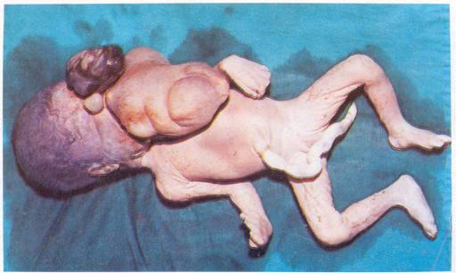
A female baby was born with masses coming out of both nostrils and the mouth (Fig. 1). Ultrasonogram at the 30th week of gestation showed a live viable fetus with a large mass around the neck and mouth. On autopsy, the mass from the right nostril showed fibrofatty tissue covered with stratified squamous epithelium. That from the left nostril consisted of brain tissue covered by stratified squamous epithelium. The large mass from the mouth was also covered with stratified squamous epithelium, and a section showed areas of hemorrhage, bone and multiple cystic spaces.
 |
|
Fig. 1. A large mass protruding from the mouth showing multiple appendages, eye like buddings and hair superiorly. Small masses are also emerging from
each nostril. |
This is a case of asymmetric conjoined twins, also referred to as "double monster",
"parasitic monster", or "parasitic twin". Asymmetric parasitic twins have one well-developed and one hypoplastic twin. If the latter is externally attached, as in this case, it is referred to as a "parasitic twin". If it is internally attached within the body of the better developed twin, it is called "fetus in fetu". Conjoined twins result from injury during the first 8-10 days after fertilization which leads to incomplete separation of blastomeres. Clinically an asymmetric parasitic twin may be similar to a teratoma; what is the differentiating point between these two remains controversial.
T.M. Anandakesavan,
I.V.G.M. Hospital,
Chalakudy,
Thrissur District,
Kerala - 680 307,
India.
