|
Ashok Saxena
Kanya Mukhopadhyay
Anil Narang
From the Division of Neonatology, Department oj
Pediatrics, Postgraduate Institute of Medical Education and Research,
Chandigarh 160012, India.
Reprint requests: Dr. Ani! Narang, Additional Professor and Head,
Neonatology Division, Department of Pediatrics, PGIMER, Chandigarh 160
012, India.
Manuscript Received: July 5,1998; Initial review completed: August 19,
1998;
Revision Accepted: October 7, 1998.
Tracheal agenesis is a rare congenital
malformation which is as yet incorrectible. Respiratory distress at birth,
absent cry, failure to intubate and successful ventilation through accidental
esophageal intubation are the hallmarks of diagnosis. We describe here a case
who had tracheal agenesis with other organ malformations.
Case Report
A male baby with a birth weight of2.65 kg was born at 34 weeks to a fourth
gravida mother by vaginal delivery. She had two nor- mal babies and had one
first trimester abortion before present pregnancy. Ultrasound during present
pregnancy showed polyhydramnios and hydronephrosis on the left side. Gestational
age by LMP was 34 weeks and by assessment was 36 weeks. The baby had respiratory
distress, cyanosis, and he did not cry at birth. Bag and mask ventilation was
started initially. Apgar at 1 minute was 3. Since there was inadequate
improvement following bag and mask ventilation, endotracheal intubation was
attempted. However, the tube could not be negotiated beyond glottis. Repeated
at- tempts at intubation failed and hence bag and mask ventilation was
continued. At one hour of life baby was "intubated" and manual IPPR was started.
Blood gases on IPPR were as follows: pH 7.37, p02 109, and pC02 33. At 3 hours
of life, the baby was spontaneously extubated and attempted intubation revealed
failure to advance the tube beyond the glottis. A diagnosis of tracheal agenesis
was entertained and esophageal intubation was done. Contrast studies were
performed by passing a feeding tube through the endotracheal (in fact an
esophageal tube). The whole bronchial tree along with feeding tube tip in
stomach (Fig. 1) was visualized bilaterally. This explained effective
ventilation through esophageal tube since both the bronchi were arising from the
esophagus. Systemic examination revealed a lump abdomen extending from left
lumbar quadrant across the midline to the right lumbar quadrant. Subsequently
the baby had severe hypoxemia and the patient expired at 29 hours of life.
Autopsy examination re- vealed: (i) Tracheal agenesis type 3 with both the
mainstenibronchi arising from the middle one third of esophagus; (ii)
Partial anomalous pulmonary venous drainage with a large ASD; and (iii)
Horse-shoe kidney with right side showing multicystic changes.
Discussion
Tracheal agenesis is a rare malformation with a reported incidence of 2 per
100000 live births(l) which was first described by Payne(2). Tracheal agenesis
is classified
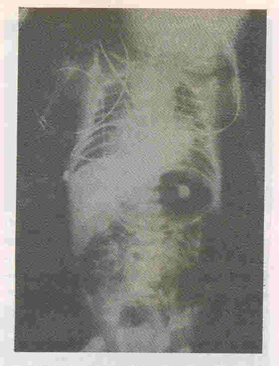
Fig. 1. Contrast X-ray shows dye in the bronchial tree and feeding tube through endotracheal tube in the stomach with gaseous distension.
according to Floyd into three types(3). In type 1 (10%), there is a normal carina and except for a short distal part, the-whole trachea is ab- sent. In type 2 (59%) the whole trachea is lacking but bronchi join in the midline. In type 3 (31 %) both bronchi arise separately from esophagus. Almost always the esophagus communicates with bronchi or trachea and this communication is called bronchoesophageal fistula. Other malformations are asso- ciated with this condition in 84% of the cases. Cardiovascular malformations are seen in 61 % of the cases while genitourinary
and gastrointestinal tract malformations occur in 40%
of the cases. As in this case absent cry, respiratory distress soon after birth and failure to
advance tube beyond glottis are the initial presenting features. Esophageal intubation, often inadvertent, results in initial improvement in ventilatory status of the baby though death is inevitable due to subsequent respiratory failure. Endoscopy of the larynx may show a blind ending pouch while esophagoscopy
will show the fistula (e) between bronchi or trachea and the esophagus. Contrast
studies visualize the fistula. Lateral X-ray of the trachea may delineate the cranial end of the trachea.
Curative surgery for this condition would consist of implantation of a tracheal prosthesis and would require preoperative ECMO. At present this approach remains in the realm of theoretical possibility. Various surgical approaches have been tried but only two cases have survived. They survived following tracheostomy. In both the cases the atretic segment was short(4).
|
|
1.
Manschot HJ, Van den Anker IN, Tibboel D. Tracheal agenesis. Anesthesia 1994; 49: 788- 790.
2.
Payne W A. Congenital absence of the trachea. Brooklyn MedicalJournal 1900; 14: 568.
3. Floyd J, Campbell DC, Dominy DE. Agenesis of the trachea. Amer Rev Resp Dis 1962; 86: 557-560.
4.
Sankaran K, Bhagirath CP, Bingham WT, Hjertaas R, Haight K. Tracheal atresia, proximal esophageal atresia, and distal tracheoesophageal fistula: Report of two cases and review of literature. Pediatrics 1983; 71: 821-823.
|