|
|
Case Reports Indian Pediatrics 2001; 38: 294-297 |
|||||||
|
Mucormycosis of the Neonatal Gastrointestinal Tract |
|||||||
|
E-Mail: [email protected] Mucormycosis is an infection caused by fungi belonging to the order Mucorales of the class Zygomycetes. They are ubiquitous organisms found in the soil and decaying organic matter. The genera causing disease in humans include Mucor, Rhizopus and Absidia(1). These fungi produce airborne spores which are the infective form. Infections occur in the rhinocerebral, respiratory, gastrointestinal or cutaneous regions depend-ing on whether the spores are inhaled, ingested or injected. Mucormycosis infection is associated with a state of immunocompromise in adults which includes diabetic ketoacidosis, lymphomas, leukemias and renal failure on peritoneal dialysis(2). The usual predisposing conditions in children are prematurity, neutropenia, acidosis and corticosteroid therapy. Mucormycosis of the gut in neonates is rare, difficult to recognize and hence usually fatal. These cases are usually diagnosed initially as necrotizing enterocolitis (NEC), which therefore leads to a delay in specific treatment. We report a case of neonatal gastrointestinal tract (GIT) mucormycosis diagnosed following laparotomy, with a review of cases of GIT mucormycosis in newborn babies from the literature.
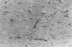
A baby boy was born to a primigravida mother at 34 weeks of gestation by normal vaginal delivery with a birth weight of 1810g and Apgar of 4 and 10 at 1 and 5 minutes, respectively. Mother had no identifiable antenatal risk factors. The baby developed respiratory distress soon after birth. Clinical features and radiological findings were suggestive of pneumonia. He was treated with parenteral fluids and antibiotics. As the baby improved, nasogastric feeds were introduced in small volume on the third day and gradually graded up. He was noticed to have abdominal distension and feed intolerance on the sixth day of life and a lower quadrant abdominal mass was felt 48 hours later. Stool for occult blood was strongly positive. His general condition deteriorated rapidly with onset of metabolic acidosis, hyponatremia and thrombocytopenia. Abdominal X-ray revealed generalized bowel loop distension without any specific radiological features of NEC including pneumatosis intestinalis. Blood culture performed twice during this period was found to be sterile. The baby was kept nil orally and treated conservatively as per the nursery protocol. However, he did not show any clinical signs of improvement and there was persistence of the abdominal mass. Thus, a laparotomy was performed on day 19. At surgery, a dense, adherent mass comprising the ileum, caecum and sigmoid colon was found, with multiple walled off perforations. A single isolated perforation was also found in the jejunum. A partial ileal resection was done and ileostomy and colostomy were fashioned. A tissue biopsy of the laparotomy specimen showed the presence of "empty-looking" broad aseptate fungal hyphae of irregular width, occasionally branching, suggestive of mucormycosis. Colonies of the fungus were seen within serosal blood vessels (Fig. 1). However, blood culture for fungus carried out at this stage was negative.
Post-operatively, the baby was provided cardiorespiratory support and IV Ampho-tericin B was commenced. In spite of these, he developed multiorgan failure with dissemi-nated intravascular coagulation and died on the 32nd postnatal day. Consent for postmortem examination was refused.
A review of literature in the English language press showed 18 case reports of neonates who presented with gastrointestinal mucor-mycosis. Majority of these (83%) were pre-mature infants. In all cases the organism was identified by histopathological examina-tion. Isolation of the organism by culture was possible in only 6(3-6) of the 18 cases (33%) and were found to belong to the genus Rhizopus. Few investigators believe that the diagnosis of mucormycosis should be made purely on histological grounds since culture may only prove presence of this ubiquitous organism and not its invasion(7). However, differentiation of species can only be made on the basis of culture. It has generally been accepted that the likely portal of entry of the organism is oropharynx, though the source of infection has been confirmed only in few cases. In one of the cases reported, Rhizopus oryzae was cultured from elastoplast. However, in cuta-neous mucormycosis, elastoplast contaminated with Rhizipus spp. was implicated as the source of infection in ten other hospitals(5). Nosocomial outbreaks of cutaneous mucor-mycosis have been reported in the hospitals where wooden tongue depressors were used as arterial splints(8). Placement of nasogastric tube has been quoted as a possible risk factor for GIT mucormycosis by causing local tissue damage and subsequent mycotic invasions. The acidic environment of the stomach may actually favor the growth of the fungus since the organism grows optimally at a pH of 6.5. In childhood gastrointestinal mucor-mycosis, the stomach is the most commonly involved organ followed by the colon, small bowel and esophagus. But in neonatal cases, the colon was predominantly involved (74%), followed by the stomach and ileum in 7 (37%) cases each, appendix in 3 (16%) cases and extra intestinal involvement in 5 (26%) cases. The clinical manifestation of majority of these patients mimicked those of NEC. However, only 3 babies presented with an abdominal mass in the initial course of the illness. It has been suggested that GIT mucor-mycosis may be a variant of NEC. The main features which differentiate GIT mucor-mycosis from NEC are the absence of pneumatosis intestinalis and poor response to chemotherapy(3). Uniformly, the prognosis has been grave with only 4 cases being diagnosed antemortem leading to surgical intervention and antifungal therapy and only 3 survived(3,5,9). Two of the babies who survived, needed a second laparotomy and surgical debridement few months after the neonatal period. The high mortality rate may be due to lack of clinical suspicion, coupled with inadequate surgical debridement and antifungal therapy. Hence, GIT mucormycosis should be suspected in any preterm baby with clinical features of NEC, absence of pneumatosis intestinalis in the abdominal X-ray and not responding to conservative management including antibacterial chemotherapy. As the diagnosis of this disease has been based on histological examination in most of the cases, a high index of suspicion with early laparo-tomy, aggressive debridement and institution of antifungal drugs would go a long way in improving the outcome of the disease. Contributors: SS drafted the paper. AKJ and RS helped in drafting the paper and analysis of collected data. ST provided the histopathological description and diagnosis. Funding:
None.
|
![]()