|
|
Original Articles Indian Pediatrics 2001; 38: 15-23 |
|||||||||||||||||||||||||||||||||||||||||||||||||||||||||||||||||||||||||||||||||||||||||||||||||||||||||||||||||||||||||||||||||||||||||||||||||||||
|
MANAGEMENT OF RETINOBLASTOMA WITH RADIATION |
|||||||||||||||||||||||||||||||||||||||||||||||||||||||||||||||||||||||||||||||||||||||||||||||||||||||||||||||||||||||||||||||||||||||||||||||||||||
|
Manuscript received: October 16, 1997; Initial review completed: December 17, 1997; Revision accepted: July 10, 2000. Objective: To evaluate the role of radiation therapy in the management of retinoblastoma. Design: Retrospective analysis. Method: From January 1993 to March 1994, one hundred and eleven children (150 eyes) of retinoblastoma were referred for radiotherapy. The diagnosis was based on clinical examination and ocular ultrasonogram for both the eyes. The radiation treatment policy involved 40 Gy in 20 fractions over 4 weeks delivered with sedation for children under 1 year of age, 36 Gy in 9 fractions over 3 weeks under ketamine anesthesia for 1-4 years of age and for >4 years of age, a dose of 50 Gy in 25 fractions over 5 weeks. The initial tumor regression was evaluated by A and B mode ultrasonography and/or CT scan. Results: The age distribution ranged from two months to six years (median - 20 months). Bilaterality was observed in 39 out of 111 cases (35%). The male to female ratio was 1.8:1. Eighty two of the 111 children were treated by definitive external beam radiation to one or both eyes. Fifteen cases received adjuvant radiotherapy after enucleation, and 14 had extensive disease for which palliative radiotherapy was offered. We observed a complete response in 54% of cases, partial response in 32%, and none in 14% of cases. Forty per cent (40%) eye survival was documented at the end of 28 months. The complication rate encountered was about 15%. Conclusion: Radiotherapy is an effective modality of treatment in significant number of patients with retinoblastoma. However, it requires appropriate fractionation, precise colimation and careful immobilization with general anesthesia.
RETINOBLASTOMA (RB) is relatively a rare form of ocular malignancy, arising from the photonuclear layer of retina. It constitutes 3% of all childhood malignancies. The incidence is however higher in the Indian subcontinent(1). The worldwide prevalence rate is 11 per million children below five years of age(2). There is a definite genetic pre-disposition to develop this malignancy. Historically, enucleation has been the treatment of choice as the ophthalmologists have been the primary treating physicians over the years. Presently, retinoblastoma is considered to be highly radiosensitive tumor. Radiation therapy (RT) is fast developing as an alternative to surgery for it offers preservation of vision and improvement in quality of life, apart from cure. Hillgartner was the first to treat retinoblastoma effectively by X-ray in 1903(3). Presently, megavoltage external beam RT and radioactive scleral plaques are being used in the management of retinoblastoma. Plaques however have a limited utility, in only selected subset of small tumors. External beam radiotherapy on the other hand can be used in all stages(4). Moreover, external RT has an advantage of treating the entire retinal lining, thereby taking care of the multicentric behavior of these tumors. Photon beam irradia-tion entails its own complications and long term sequlae. Several centers, therefore, are utilizing other modalities such as cryotherapy and photocoagulation in suitable patients(5,6). We studied 111 patients of RB to establish the role of radiotherapy as a primary modality of treatment in all stages of RB, including bilateral and locally advanced disease.
Patient Selection One hundred and eleven patients referred from Rajendra Prasad Eye Center were registered in the Department of Radiation Oncology, Institute Rotary Cancer Hospital, All India Institute of Medical Sciences, between January 1993 to March 1994. The clinical and family history, and systemic examination finding were recorded in all cases in collaboration with an ophthalmologist, and a pediatric oncologist. Fifteen patients were previously enucleated and required adjuvant postoperative radiotherapy. Investigations Detailed ophthalmological examination included indirect ophthalmoscopy (IO), examination under anesthesia (EUA), visual field charting and visual acquity test. Routine investigations like complete blood counts, liver, and kidney functions were carried out for systemic evaluation. In advanced cases, metastatic workup in the form of bone marrow biopsy, CSF cytology for malignant cells, and bone scan were done. The intraocular tumor volume estimation was done by orbital A and B scan and/or a CT scan. Treatment Protocol Eighty two patients were considered for radical radiotherapy, 15 for adjuvant post-operative RT, and 14 for palliative RT. Patients considered for radical RT were treated age wise by different protocols. Children below 1 year were treated with 40 Gy in 20 fractions over 4 weeks (200 cGy per fraction). Children between the ages 1 to 4 years received 36 Gy in 9 fractions over 3 weeks (400 cGy per fraction), and for children over 4 years, a dose of 50 Gy in 25 fractions (200 cGy per fraction) was delivered. Children less than 1 year were treated under sedation, and those less than 4 years were immobilized under ketamine anesthesia. The radiation was delivered by 1,2 or 3 field technique, depending upon the stage of the disease, by a telecobalt unit. A single temporal port was used for early stage (<10 mm tumor height) unilateral disease. For early bilateral tumors lateral portals were designed on either side, with 3-5 degrees posterior tilt, in order to avoid exposure to the contralateral lens. In locally advanced unilateral disease, lens had to be included, and so radiation was delivered by two fields anterolateral portals in order to cover the entire mass. Here again, a posterior tilt of 10 to 15 degrees was given to the lateral field, so as to avoid irradiation to the opposite lens. In case there was asymmetric distribution of the tumor in both the eyes, the irradiation technique was similar to their respective tumor bulk in each eye, but total dose prescription was made at middle after considering the contribution of the doses made from each beam to each eye. When there was bulky disease in both the eyes, a single anterior portal was added to the temporal ports, thereby sacrificing the sight. Assessment and Follow-up The tumor regression was assessed both by clinical examination and ultrasonography at every month till first-2 years and thereafter every 4-6 months. Patients of phthysis bulbi were assessed by CT scan at 4 to 6 monthly interval. In patients who had progressive disease, salvage modalities of chemotherapy and/or surgery (enucleation/exenteration) were undertaken. Statistical Analysis Kaplan-Meier method was used for estima-tion of overall disease free survival (DFS)(7).
The age distribution of these patients varied between 2 months to 6 years with a median age of 20 months. The male to female ratio was 1.8 : 1. One hundred and fifty eyes were found involved by retinoblastoma out of which 72 (65%) cases had uniocular disease. The tumor volume estimation from EUA finding, orbital USG scan and CT scan showed early intraocular disease (<6 mm) in 14 cases, moderately bulky intraocular disease (6-10 mm) in 12 cases. Majority of the cases (n = 53) however, had locally advanced disease, i.e., tumor height greater than 10 mm or upto back of the lens. Vitreous seeding, which too was included in this group, was seen in 51 cases. Extraorbital mass lesion was seen in 19 cases (Table I). Fifteen cases received RT in adjuvant setting post-operatively and 14 received palliative RT, either locally (n = 11) or to a distant metastatic site(s)(3).
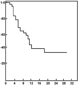
After one month of completion of RT, fifty-four per cent cases had complete regression with classical "cottage cheese appearance" on the USG scan. Thirty two per cent of all these patients who were treated by radiotherapy had a partial response. In all these cases it was difficult to assess the viability of the residual mass. Sixteen (14%) cases however, failed to show any response. Minimal and maximal follow-up duration ranged between 1 to 33 months (median follow-up 11 months). The eye survival was recorded as 40% at the end of 28 months. The median disease free survival period was 11 months (Fig. 1). The complications document-ed were mild to moderate phthysis bulbi, red eye and lid swelling. The total complication rate was 15%.
Failure Pattern The maximum failure rate was encoun-tered between 6-16 months post treatment after which plateuing of response was observed. Of the fifteen postoperative cases treated, one failed at 12 months at the local site, and three developed distant metastasis at 18, 10 and 8 months. Amongst the patients who received palliative radio-therapy, the objective response was good (in 70% cases) at the treated site. Of the 111 cases a total of 12 cases developed distant metastases in CNS, scalp, or bone during the course of follow-up; of which one responded to treat-ment and four succumbed to disease. Bilateral retinoblastoma was observed in 39 cases and of these most were synchronously bilateral, and only 4 showed metachronous disease. Amongst the bilateral 3 cases showed strong hereditary link (family history positively). Family history was found to be positive in 7 cases in the entire series.
In the treatment of RB the two aims are cure and preservation of vision. This is possible, with radiotherapy, only by inclusion of entire retina and protection of lens. The disease specturm encountered in a referral center like ours is that of advanced nature where preservation of vision may not be always possible (Fig. 2). This picture however, is not seen in the western literature(8). In this study, we encountered a slight male pre-ponderance unlike the studies reported earlier where no sex predilection was observed(3). Family history was positive in 7 cases, in the entire series, of which 3 were bilateral. Such a low familial incidence is possibly due to lack of many long survivors of previously treated retinoblastoms, and due to lack or awareness amongst the parents and caretakers. Our study revealed bilaterality of 35% which is consistent with the literature(1,8,9). There was no right or left eye predilection. Leukocoria which is the commonest presenta-tion was seen in 70% cases and this is in consensus with other studies(10). A and B mode ultrasonography was carried out in all the patients with intraocular disease. This has proven to be a very useful diagnostic tool in retinoblastomas as observed in this study and an earlier report from our institution(11). Not only is it reliable for differentiating this tumor from other pathologies which simulate its presentation, but it also has an established role during the follow-up period. Plain radio-graphic studies are no longer being recom-mended as the calcification is encountered in variable number of cases ranging from 20% to 80%(12). CT scan is definitely preferred in these tumors, especially since the advent of high resolution scanners (Fig. 3). MRI scan-ning is not as specific as the above mentioned investigative tool as it is unable to detect calcification, but due to its high resolution it is definitely superior for assessing the optic nerve involvement(13).
Since early parts of the century, it has been established that radiation could effectively destroy retinoblastoma(14). Several workers now feel that enucleation should no longer be recommended in a unilateral case for it may become bilateral at a later date. And, contrary to the earlier belief that in bilateral disease the worse affected eye should be enucleated and the better eye irradiated, it is now possible to effectively irradiate both eyes even in cases of uneven distribution of tumor bulk(15). At our center the patients underwent external beam radiotherapy with an intent to cure by irradiation. Patients who received radical treatment were irradiated either by lens sparing technique or by anterolateral portals with differential loading towards the anterior portal, in cases where vision preservation could not be contemplated. Bilateral tumors were treated by two parallel opposed portal with or without addition of an anterior field (to the bulkier tumor). Although, several staging systems have been in use for retinoblastomas, for radiotherapeutic approaches, Reese-Ellsworth grouping system is considered suitable(16) to compare between different series. Since a staging system should reflect natural history of the tumor, and discriminate patient groups with differing prognostic features, Reese-Ellsworth staging system seems inadequate in present times. Howarth et al.(8) have devised their own system where four stages of tumor involvement are recognized: (i) retinal, (ii) intra-ocular, (iii) regional, and (iv) metastatic. We have further attempted to clinically stage this tumor for the sake of convenience at the time of decision making regarding treatment. This staging takes into consideration that multimodal approach is essential in tackling different stages of the tumor. The main thrust of this staging system is on the tumor bulk as outlined in Table I. We followed the standard high dose per fraction schedule using 36 Gy in 9 fraction over 3 weeks in the children who needed anesthetic immobilization(17). Foote et al. (18) have reported that a dose of 45 Gy in 1.8 Gy fractions appears to be adequate for local control of tumors smaller than 10 disc diameters. Larger tumors may however require higher doses. Since most of our tumors were bulky, except for children below 1 year of age, we have delivered the dose equivalent to 50 Gy in all other patients. In bulky tumors, the main difficulty in the irradiation of retinoblastoma has been to deliver a high uniform dose to the entire retinal surface. We recommend the anterolateral portal which is reproducible and can be carried out with relative ease. In cases of antero-lateral portals a posterior angulation of 10-15° to the lateral beam was considered optimal, although some authors recommend an angulation of up to 40° in posteriorly situated tumor to spare the opposite retina(19). Chin et al.(20), however, recommend a non-coplanar technique for such tumors, which gives higher doses to the ora serrata. Sandars et al.(21) report a 3 year survival rate of 90% when all modalities of treatment such as surgery, radiotherapy, chemotherapy, cryotherapy and photocoagulation were used. Similarly another large series reported by McCormic et al.(22) achieved 4 year survival of 93% (after salvage). Cassady et al.(23) however, in 1969 reported a local control for all stages of disease as 49% with radiation alone which rose to 73% in patients with group I to III disease (Reese-Ellsworth staging). Our survival rates with radiotherapy are also less when compared to other studies(24,27). In our series the overall local control rate was 40% at 28 months (Table II). We encountered 15% complication rate of which phthysis bulbi was the commonest. Howarth et al.(8) suggest that extraction and use of corrective lenses may provide good vision, but in our experience isolated cataract formation without phthysis development and corneal opacification was not seen; hence such rectification procedures would not be possible. No second malignant neoplasms have been observed so far in this group of children with retinoblastoma, but survivors of this tumor should be observed for the same, for longer periods of time as studies have reported upto 10% incidence of second malignancy in a long term follow-up(26).
The median interval from diagnosis of intraocular retinoblastoma to the development of orbital recurrence was 6 months which seems to be the same as the period following enucleation(27). Apart from intraorbital relapse, spread to the brain parenchyma is observed occasionally. The other sites of relapse are CSF, bone, lung(23,28). Death was documented in 4 patients in our series (out of 12 with distant spread). Four cases were salvaged by surgery and 11 by chemotherapy. Prognosis however remained poor in them. The mortality as reported in the literature is approximately 9%(3). The possible explanation for the poor results in this series are – late presentation of the disease, large number of cases with vitreous seeding which can remain as viable and hypoxic cells following irradiation. Early disease was encountered unilaterally in 12 cases, of which 5 cases were free of disease at the end of follow up. There were 4 uniformly early bilateral cases, of which 3 do not have any disease at the end of follow-up. Of the 39 bilateral cases, 18 had unequal disease where one eye had an early tumor, and no difference in the outcome was seen in these cases. It is, therefore, inferred that bilaterality did not affect the prognostic value of these tumors, but the individual tumor burden did(15). These tumors showed inherently aggressive bio-logical behavior and therefore presented with bulky disease early in the natural course of disease. This could possibly be explained by a tendency to early retrolaminar and choroidal invasion(28) and optic nerve involvement(29). We suggest studies would have to be carried out in order to explore the exact reason as to why the retinoblastoma load is higher in India(1) as comapred to other countries and, that the retinoblastoma clone in India does not respond as well to radiation treatment as reported in Western literature. In conclusion, radiotherapy requires fractionated treatment, precise collimation and localization, careful immobilization with anes-thesia and a prolonged observation period for evaluation for control of tumor and complica-tions of therapy(26). Moreover, we would like to suggest that larger tumor burden (occupying the major portion of posterior chamber) may not exactly be well taken care of by radiation alone. Therefore, a judicious use of investigative tools such CT scan, MR scan etc. along with the use of treatment modalities such as chemotherapy in extraorbital tumors and cryosurgery in smaller masses(6) may be required to enhance the results of this tumor. Contributors: PL and BMB evaluated the patients for radiotherapy and drafted the manuscript., BKM and GKR supervised the radiotherapeutic management of all the patients. DNS performed literature search and helped in drafting the manuscript; he will act as the guarantor for the study. SG carried out the ophthal-mic assessment and management. TV and LSA administered chemotherapy and looked after the other problems encountered during treatment. Competing interests:
None stated.
| |||||||||||||||||||||||||||||||||||||||||||||||||||||||||||||||||||||||||||||||||||||||||||||||||||||||||||||||||||||||||||||||||||||||||||||||||||||
![]()