|
|
Case Reports Indian Pediatrics 2000;37: 1370-1373 |
|||||||
|
Bilateral Adrenal Cysts in a Newborn |
|||||||
|
Cystic lesions of the adrenal gland are relatively uncommon, with the reported incidence of 0.06-0.18%(1). As most lesions are small and asymptomatic, true incidence of adrenal cyst is not known. Adrenal cyst occurs in all age groups but are relatively rare in children. Adrenal cyst is usually unilateral and is most commonly seen on right side(2). However, in our case it was present on both sides with the left side being larger in size, which prompted us to report this neonate.
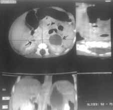
A two-day-old female neonate was admitted in our hospital with complaints of poor feeding, soon after birth. The child was a full term appropriate for gestational age (2.6 kg), product of non-consanguineous marriage, born by normal vaginal delivery at a private hospital. Antenatal period was uneventful but the baby was delivered after a precipitate labor. At the time of admission, on day two of life, general physical examination revealed a lethargic newborn with poor cry and activity, circumoral cyanosis and vacant look. Rest of general and systemic examination was normal. Blood sugar was found to be 20-40 mg/dl by dextrostix and was confirmed to be less than 25 mg/dl by conventional method. Serum calcium was normal. The baby was adminis-tered intravenous fluids with 8 mg/kg of glucose per minute. Since sepsis screen could not be ruled out on admission, the baby was given intravenous antibiotics. Blood sugar was continuously monitored using dextrostix. Over the next 3 days the baby required glucose infusion as high as 12 mg/kg/min for which umbilical vein had to be cannulated. Urine was negative for reducing substances. X-ray abdomen was normal. Ultrasound abdomen was performed to rule out any pancreatic pathology but surprisingly an ill defined hypoechoic mass was found in the left suprarenal gland. Serum cortisol level couldn’t be done due to financial constrains. Tube feeds were started from day 3 of life and intravenous fluids were gradually tapered till the baby accepted full feeds. The baby was discharged after staying for 11 days in hospital. CT scan abdomen done in the follow up revealed a nonenhancing hypodense area, with rim calcification, of the size of 7 ´ 2 ´ 2 cm in the left suprarenal gland and similar area of approximate 1.5 cm ´ 1 cm ´ 4 mm size in the right suprarenal gland (Fig. 1). These findings were suggestive of bilateral adrenal cysts, probably hemorrhagic in nature. Repeat ultrasound abdomen done at 4 months of age showed complete resolution of the adrenal cyst. The child has been growing normally on follow up.
Adrenal cysts are rare though their true incidence is probably higher than reported(1). This observation in due to the more frequent diagnosis of asymptomatic incidental cysts noted on imaging studies(3). Adrenal cysts may occur at any age, but most are found in the 3rd through 4th decades of life(4). In some series, a female preponderance of about 3 : 1 has been noted for unknown reason(5). There is a paucity of publications of adrenal cysts in newborns. Adrenal cysts are usually asymptomatic and many are discovered at autospy. Symptoms, if present, are related to size and position of the lesion and can include pain, gastrointestinal disturbances, or the finding of a palpable mass. They seldom cause adrenal hypofunction(6), Cushing’s syndrome(7), or pheochromocytoma(8). Patients may present with acute abdominal findings if intracystic hemmorhage or rupture occurs(9). In our case the probable cause of hypoglyceymia was adrenal insufficiency, though we could not confirm it. Adrenal cysts can be divided into four groups(5); (i) Endothelial cysts (45%); (ii) Pseudocysts (39%); (iii) epithelial cysts (9%); and (iv) Parasitic cysts (7%) Pseudocysts are the second most common type of adrenal cyst but are probably the most common clinically recognized type of cyst(4). These are usually large and uniloculated. The walls are composed of dense fibrous connective tissue with islands of adrenal cortical tissue incorporated. Pseudocysts may occur as a result of hemorrhage within normal adrenal tissue or within a tumor. Pseudocysts have been reported in children following adrenal hemorrhage as a result of injury or infarction in the neonatal period(10). Our case belonged to this category of adrenal cyst. Adrenal cysts may get complicated by getting infected or by compressing on adjoining structures. Function-ing adrenal cysts are those which have islands of adrenal cortex incorporated into cyst wall and have clinical and biochemical evidence of hyperadrenalism. Diagnosis may be suggested by a soft tissue swelling with curvilinear calcification seen in Plain X-ray abdominal films. Ultra-sonography and CT scans are useful diagnostic tools and many adrenal masses are discovered incidentally. Treatment of adrenal cysts is determined by size and symptoms related to the mass. Conservative management is appropriate for newborns with adrenal hemmorhage and resulting pseudocyst formation. Spontaneous resolution of pseudocysts in newborns usually occurs by 6 weeks, and some reports support waiting for as long as 6 months prior to planning intervention(11). We followed up our patient conservatively and the cyst resolved spontaneously. Other varieties of adrenal cyst may require intervention due to complica-tions. Percutaneous aspiration or drainage may be an acceptable initial management strategy(12). Surgical excision also can be done for large and complicated cyst as well as for parasitic, functioning and malignant cysts. Contributors: BB and RK were involved in diagnos-ing and investigating the case under supervision of AKP and VKA. BB drafted the paper which was edited by AKP. Funding:
None.
|
![]()