|
|
Personal Practice Indian Pediatrics 2001; 38: 995-1008 |
|||||||||||||||||||||||||||||||||||||||||||||||||||||||||||||||||
|
Three Common Dermatological Disorders in Children (Scabies, Pediculosis and Dermatophytoses) |
|||||||||||||||||||||||||||||||||||||||||||||||||||||||||||||||||
|
As nearly 30% of all outpatient visits to a pediatrician consist of either primary or secon-dary dermatological cases(1), a knowledge of some of the common dermatological problems is important. Two cutaneous infestations, namely scabies and pediculosis and superficial fungal infections (ringworm) are most commonly seen, yet often treated inappro-priately. This section discusses how to easily diagnose and manage such patients as these three conditions are easily amenable to therapy. Scabies Scabies or "the classical itch" caused by the human mite, Sarcoptes scabeii var hominis is the most common ectoparasitic dermatosis seen in clinical practice. Although not a life threatening condition, yet it may be considered to be important from the public health point of view because of several reasons, namely it is found globally; severe itching is extremely distressing and the impetiginized cases may be complicated by post-streptococcal glomeru-lonephritis. By systematic approach this condition can be managed appropriately and its transmission can be prevented in the community(2). Etiology The human scabies mite, ever since its rediscovery and establishment as the causative agent of scabies in 1687 by Bonomo and Cestoni, has remained a challenge as far as an ideal and safe scabicidal agent is concerned(3). The female mite which is bigger than the male; has four pairs of legs and a rounded body (400 µm in length), has a life-cycle of 30 days, during which it lays 11 to 25 eggs(3). It burrows through the stratum corneum of the skin and the itching is due to a type-IV hypersensitivity reaction in the skin to the mite and their secretions(2). After an incubation period ranging from 3 to 4 weeks, the patient presents with intense itching which is worse at night(4). Epidemiology Scabies affects hundreds of millions of people worldwide(5). The highest prevalence rate may be seen in children, especially below 2 years of age(6). It is truly a disease of the rural tropics because it is a disease of poverty(2). The point prevalence in the general population of rural communities in India is about 5%(7). Both sexes are equally involved. The important predisposing factors are overcrowding, poor hygiene, population shifts, immigrations and sexual contact. The source of transmission is by close personal contact within the household. Fomites are relatively unimportant sources of transmission, as demonstrated by some authors(8). Diagnosis and Clinical Features If approached systematically, the diagnosis of scabies can hardly be missed. An easy method to diagnose a case of scabies is given in Table I.
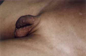
Variants of Scabies Nodular Scabies: Sometimes, itchy nodules may persist for several months and are found most commonly on the scrotum (Fig.4). Scabies incognito: Due to local application of corticosteroids, scabies becomes modified and this form is known as scabies incognito. Norwegian scabies: A rare form of scabies can occur in patients who are mentally retarded or immunocompromised, including those with HIV/AIDS, which consists of hyperkeratotic and crusted lesions on the hands and feet. It is a highly infections condition, since there are innumerable mites in the scales(2,3). Animal or canine scabies: Various itch mites affect dogs, cats and birds and these can be acquired by human beings on close contact with these animals(9,10). Dogs are the major source(10). Clinically, canine scabies trans-mitted to humans looks different from that produced by the human itch mite. An itchy, papular eruption is localized to site of contact of the animal with the human skin. Burrows are conspicuous by their absence. As the canine mite is unable to complete its life-cycle on the accidental human host, once the animal is treated with antiscabetic agents or removed from the environment, the human infestation resolves(3). Only symptomatic treatment should be given to the child.
Fig. 1. Sites of involvement in scabies in infants and children.
Fig. 2. Sites of involvement in scabies in adults. Demonstration of the Mite A scraping of papule or a burrow can be done to confirm the clinical diagnosis. Mineral oil is placed on a number 15 scalpel blade, which is used to scrape several lesions. The scrapings are transferred to a glass slide. Under the light microscope, one may then look for mite, eggs or fecal pellets known as scybala(2,5). However, the history and clinical examination are adequate to make the diagnosis. Treatment Although the often emphasized and seemingly simple single application treatment of scabies with scabicides, seems to be a natural solution, in reality, perhaps no other treatment has been as much misunderstood as that of scabies. Table II gives the general instructions and the correct method to apply a scabicidal agent and Table III summarizes the commonly used scabicidal agents in children. It must be emphasized that all these topical agents are effective for treating scabies in children, if used correctly and each has its advantages and disadvantages. According to a recent Cochrane review, the conclusion is that 5% permethrin cream is probably the most effective and safest treatment for scabies in children beyond two months of age(11), where sulphur and crotamiton cream are safer yet less effective choices(5,6). Ivermectin, used systemically holds promise for the future as it has been shown to be effective in children by some authors although its safety profile is still to be determined in more randomized controlled trials(5,6,13). It is mainly approved for use in children 5 years of age and above(6). Clinical data of usage in children below 5 years of age is lacking. Table II - General Instruction to the parents of a child with scabies
Table III - Commonly used scabicidal agents in children (2,5,11-13)
Treatment of secondary infections with antibiotics is necessary. The proper treatment of presenting cases and their contacts during epidemics and localized outbreak is a good approach. Thus, scabies which may appear to be an innocuous dermatosis in children, could prove to be a major burden to public health if not managed adequately. Pediculosis Pediculosis capitis is caused by the head louse Pediculus humanus capitis; pediculosis corporis by the body louse, Pediculus humanus corporis; and pediculosis pubis by the pubic or crab louse, Pthirus pubis. They are obligate parasites of man and highly host specific. The entire life-cycle is completed in 1 month(14). The lice are dorsoventrally flattened wingless insects witht the body divided into 3 segments and have 3 short pairs of legs. Epidemiology Pediculus humanus capitis is distributed worldwide. It is transmitted by hair-to-hair contact and by fomites (e.g., hair accessories, hairstyling tools and clothing). It attacks all, transcending social status. Healthy children between the ages of 3 and 10 years are primarily affected(6). Girls may be affected more than the boys. Pthirus pubis infestation occurs mainly in adolescents and adults and is spread sexually or by fomites such as bed linens and towels. It may also involve other areas where short hairs are found such as the trunk, extremities, axillae, beard, moustache, eye-brows, eyelashes and occiput. Pediculosis corporis also occurs worldwide and affects the homeless and underpriviledged. Clinical Features Pediculosis Capitis It is quite simple to make a diagnosis of pediculosis capitis and an approach is given in Table IV.
Management The treatment for pediculosis capitis(5,6,14-18) is given in Table V. Standard therapy includes the topical application of pediculocides(5,6,17,18). The treatment of choice is either permethrin or lindane in children although other alternative therapies can be tried. Two applications, a week apart are recommended as none of the available treatments are completely ovicidal(2,5). After treatment, nits may be removed either by dry combing(15) or soaking the hair with white vinegar (3% to 5% acetic acid) or 8% formic acid rinses to soften the cement of nits before combing the hair. Physical modalities such as mechanical nit and louse removal can also be attempted. This is important because ideally the child should be nit free before returning to school. Fomites may be washed in soap and hot water or stored in a sealed container. Secondary pyodermas must be treated and oral cotrimoxazole is often effective in controlling the pyoderma as well as killing the adult lice. Table V - Pediculocides used for treatment of pediculosis capititis in children
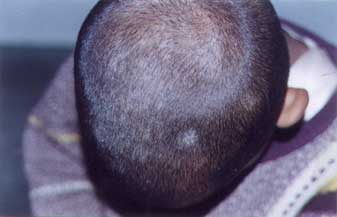
Pediculosis Corporis Nocturnal itching is the main symptom. The body lice, Pediculus humanus corporis are found in the clothes’ seams and the nits are firmly attached to cloth fibers. Small erythe-matous papules occur on covered areas of the body such as the trunk, axillae and groins. Excoriations, postinflammatory hyper- pigmentation, weals, secondary bacterial infections can also occur. Simple hygienic methods, including bathing the patient and washing and ironing all the clothes and linen is necessary. Although treatment of the body is not required, a single application with a pediculocide may prove to be helpful at times(5). Clothes can be dusted with 10% DDT, 10% gamabenzene hexa-chloride or 0.5% malathion dusting powder. Pediculosis Pubis The patient presents with itching of the pubic region, although other areas such as eyelashes, eyebrows, axillae, perianal area, beard and scalp can also be affected. In adults, the disease is contracted chiefly as a result of sexual intercourse and in a child, eyelash infestation maybe a marker of child sexual abuse(6,9). On examination, small brown lice may be found attached to the base of hairs and nits can be found attached to hair shafts. The pathognomic lesions are maculae cerulae, blue or slate-gray 0.5-1 cm macules, which are caused by salivary enzymatic degradation of bibrubin to biliverdin(8,19) and are occasionally seen in fair skinned individuals. For treatment, any of the pediculocides can be used. The whole body should be treated and a repeat application can be given after 1 week. Shaving of hairs is another alternative treatment. For eyelash infestation, petrolatum applied 3 to 5 times a day, can asphyxiate lice and nits(3,20). Besides this, 10-20% fluorescin or manual removal of lice with forceps can be attempted. Linens and clothings should be washed. Dermatophytosis Dermatophytosis (tinea or ringworm) is caused by dermatophytes of species Tricho-phyton, Microsporum and Epidermophyton. The dermatophytes are superficial fungi which thrive only on keratin layer and do not invade the living epidermis. Ringworm is classified according to the name of the affected part; these nomenclature include tinea capitis (scalp), tinea corporis (body), tinea cruris (groins and upper thighs), tinea pedis (foot), tinea barbae (beard and moustache area), tinea faciei (glabrous skin of the face), tinea manuum (hands) and tinea unguium (nails). The dermatophytes can be grouped according to the ecological headquarters like geophilic species (origin from the soil); zoo-philic species (animal origin) and anthrophilic species (confined to human skin). Anthrophilic species cause mild but chronic disease, however, zoophilic and geophilic species produce inflammatory reaction in man. Epidemiology Dermatophytosis is found worldwide and accounts for 8 to 10 per cent of skin referrals. Etiologically, Trichophyton rubrum is the commonest agent causing dermatophytic infection(21,23). The commonest dermato-phytosis in children is tinea capitis seen mainly in urban pre-school aged and school aged children(24-27). In southern India, 60 to 80 per cent cases of tinea capitis is caused by Trichophyton violaceum (28,29). Tinea pedis, manuum, corporis, barbae and cruris occur mainly in adolescents and adults. The mode of spread of infection is by contact with an infected person or animal. Fomites such as infected combs, brushes, caps, hats, couches, floors of swimming baths also transmit infection. Tinea Capitis Tinea capitis occurs almost exclusively in children. After puberty, its occurrence is rare due to inhibitory effect of secretions by sebaceous glands(9). It may be broadly categorized into two types, namely, non-inflammatory and inflammatory tinea capitis. When in invades the scalp stratum corneum and the hair cuticle. it is called ectothrix infection and when the fungus invades the interior of the hair shaft, it is called endothrix(23,30). Non-inflammatory Tinea Capitis: This is characterized by partial or complete alopeciac areas of oval or round shape with minimal inflammatory features. Black dot type: In this, there are discreate areas of alopecia studded with black dots, representing broken off hair, It results from endothrix infection of the hair. Grey patch: In this variety there are scaly, alopeciac patches with dull, grey hairs broken of at different lengths (Fig. 5). This is due to ectothrix infection of the hair. Seborrheic dermatitis like: Here the picture of the scalp resembles that of dandruff with increased, dry, scaling and less visible alopecia. Inflammatory Tinea Capitis These varieties of tinea capitis heal with eventual scarring.
Tinea Corporis and Faciei The characteristic lesions consist of single to multiple, small to large, itchy, erythematous, well defined circinate or circular lesions with the border studded with papules, vesicles, pustules or crusts and a central clearing (Fig.7). Tinea Cruris This occurs mainly in adolescents or adults wearing tight fitting garments. The groins, thigh, buttocks and scrotum are affected and the lesions are bilaterally symmetrical, itchy, well marginated, erythematous and scaly. Tinea Pedis and Manuum In tinea manuum, the lesions are usually unilateral and appear as pruritic, diffuse hyperkerkeratosis with fine, white scales or circular, scaly patches on the palm. Tinea pedis may appear as itchy maceration between the toes or diffuse hyperkeratosis with scales on the soles and heels or a vesicular type with painful blisters.
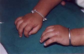
Fig. 3. Excoriated erythematous papular lesions of scabies on hands and forearms.
Fig. 4. Persistent scabies nodules on male genitalia.
Fig. 5. Grey patch variety of Tinea capitis
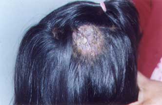
Fig. 6. Kerion
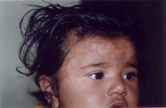
Fig. 7. Tinea faciei–erythematous plaque lesion with central clearing and border with papules and crusts.
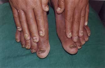
Fig. 8. Tinea unguium–discoloration, thickening and crumbling of nail. Tinea Unguium There is infection of the distal and lateral nail plate with discoloration, thickening, and crumbling of nail (Fig.8). Onycholysis and subungual keratotic debris are seen at the affected site of the nail plate. besides this, a superficial type involving the nail surface may also occur. All nails are seldom affected and full nails are never destroyed. Diagnosis The infections can be confirmed by microscopic examination of the skin scrapings taken from the surface and border of the lesions with a blunt scalpel or hair plucked with forceps or nail clippings or subungual debris(23,25). A drop or two of 10 per cent KOH are added to the scrapings on a glass slide, covered with a cover slip and allowed to stand for 10 to 15 minutes for the dissolution of keratin. Hairs and nails require a longer time. These are then viewed under the microscope for mycelia and spores. Culture can be done on Saboraud’s agar or Dermatophytic Test Medium (DTM). In tinea capitis, brilliant green fluorescence is seen on Wood’s light examination of the affected hair. Treatment Topical therapy in the form of any of the available antifungal agents (creams, lotions, powders) of imidazoles (1% clotrimazole, 2% miconazole, 1% econazole, 2% ketoconazole); 1% tolnaftate, 1% ciclopiroxolamine, Whit-field’s ointment (6% benzoic acid and 3% salicylic acid) is given for two to four weeks or continued for a couple of weeks after clinical subsidence. Systemic therapy is given for extensive infections, tinea capitis, tinea unguium, tinea manuum and pedis. The standard therapy of choice is microsized griseofulvin which is given orally in a dose of 10 to 20 mg/kg/day in two divided doses with a fatty meal for two to four weeks for tinea cruris, corporis and faciei, four to six week for tinea capitis; six to eight weeks for tinea pedis and manuum, six months for fingernails infections and six to twelve months for toe nail infections. Other systemic antifungals such as ketoconazole, fluconazole and the newer antifungals, terbinafine or itraconazole have also been tried(31,32). An effective second line agent may be required in instances when griseofulvin cannot be tolerated or there is poor response to griseofulvin therapy due to griseofulvin resistance and for tinea capiti, which may frequently require longer therapy and tinea unguium which often responds poorly to griseofulvin. Itraconazole, in a dose of 5 mg/kg/day orally has been tried in two to three monthly pulses of one week for tinea unguium in children by some authors(32,33) and shown good results. Itraconazole has also been tried in a continuous dose of 100 mg/day for 6 weeks for tinea capitis in children and compared to griseofulvin, identical cure rates(88%) were achieved(34). Studies have demonstrated that both continuous as well as pulse therapy have been found to be safe and well tolerated in children(34,35). Moreover, no relapses occurred after clinical cure at one to two years after pulse itraconazole therapy in children, making it a better alternative to griseofulvin(35). Terbinafine, has also been shown to be very effective (93% cured) in the treatment of children with tinea capitis, using a shorter treatment duration ( 4 weeks) than that usually employed with presently available antifungal drugs including griseofulvin (31,32,36). Its use has not been documented in children less than 2 years of age (less than 12 kg). The dose of terbinafine is 62.5 mg/day in children weighing less than 20 kg, and 125 mg/day for children weighing between 20-40 kg while for children weighing more than 40 kg, the adult dose of 250 mg is appropriate (36,37). The recommended duration of treatment in children is 4 weeks for tinea capitis, 2 weeks for tinea corporis and pedis, 6 weeks for finger-nail onychomycosis and 12 weeks for toe-nail onychomycosis (36,37). In a pilot study of use of terbinafine in tinea capitis in children, no relapse were observed 2 weeks after completion of treatment(37). Thus although, both itraconazole and terbinafine appear to be more promising in childhood dermatophyte infections than griseofulvin, as they have shown higher clinical cure rates, clinical safety, and lower rates of relapse, the high cost of both is the limiting factor in a country like India, where griseofulvin still remains the mainstay of treatment in childhood dermatophytoses. Other Measures For tinea capitis, washing of the scalp with selenium sulfide 2.5% shampoo, twice weekly is recommended(5) to prevent transmission by fomites. For treatment of kerion, 1mg/kg of predisolone may also have to be added initially. Combs, brushes, towels, clothes, swimming pools should not be shared by infected persons. Children being treated for tinea capitis may attend school and shaving of head or haircuts are not required. Contributors: RS reviewed the literature and drafted the manuscript. AJK provided the concept and criti-cally revised the article; he will act as the guarantor for the manuscript. Funding: None Competing interests: None stated.
| |||||||||||||||||||||||||||||||||||||||||||||||||||||||||||||||||
| Reference | |||||||||||||||||||||||||||||||||||||||||||||||||||||||||||||||||
|
|
![]()