|
|
Images in Clinical Practice Indian Pediatrics 2000;37: 1021-1022 |
|
Diaphyseal Aclasia (Multiple Exostoses) |
|
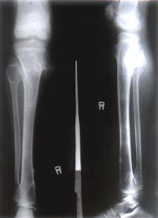
Skeletal survey showed multiple osteo-cartilagenous tumors at the metaphysis of the distal femur, proximal and distal tibia and fibula; (Fig. 2) distal radii and ulna, proximal phalanx and of right middle finger with widening of metaphysis. These changes are consistent with sessile and pedunculated multiple diaphyseal aclasia. Multiple exostoses is an autosomal domi-nant abormality commoner in males. Cartilage capped osteomas occur at the metaphysis, particularly at the ends of the long bones. These osteochondromes grow slowly with the child, cease to grow at maturity and may cause local problems by interfering with adjacent structures. A policy of selective excision should be adopted. Malignant changes may occur in adult life, particularly with exostoses affecting flat bones. Any growth in an exostoses after skeletal maturity should be considered suspicious of malignancy. Sutapa Ganguly,
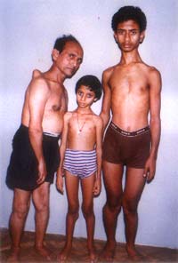
Fig. 1. The girl with her father and brother. Multiple bony projections seen around knee joints, on the ribs and on the acromion process of right scapula of the father.
Fig. 2. X-ray of the girl showing multiple osteo-cartilagenous tumors at the metaphysis of distal femur and promixal and distal tibia and fibula. |
![]()