There is still a dearth of data of the involvement of
skin in the coronavirus disease 2019 (COVID-19), especially in pediatric
patients. Herein, we describe the report of a child with COVID-19
related multisystem inflammatory syndrome in children (MIS-C), who
developed hypocomplementemic urticarial vasculitis syndrome (HUVS) after
recovery.
A previously healthy 18-month-old boy with a five day
history of fever and abdominal tenderness was admitted to the pediatric
in-patient department. His mother was suffering from COVID-19. On
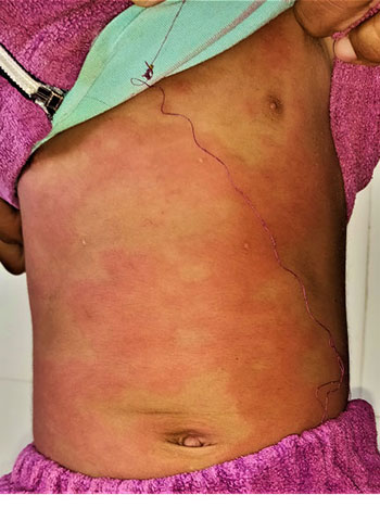
mucocutaneous examination, the child had multiple annular polycyclic
erythematous plaques on trunk with conjuctival erythema (Fig. 1).
The lesions had been rapidly progressive and persistent for the last
three days. The child was febrile (39.4°C) and hypoxemic. The child was
also experiencing diarrhea for three days along with hypotension (blood
pressure 90/60 mmHg). Laboratory investigations revealed a positive
RT-PCR for SARS-CoV-2 on two tests done three days apart, along with
metabolic acidosis, leukocytosis, neutrophilia, lymphopenia, anemia and
hypoalbuminemia with albuminuria. Erythrocyte sedimentation rate (21 mm
first hour reading) and C-reactive protein (19 mg/dL) were raised.
 |
|
Fig. 1 Multiple annular polycyclic
erythematous urticarial plaques on trunk.
|
High resolution computed tomography (HRCT) showed
ground glass opacities in <20% of both lungs. A diagnosis of MIS-C was
made. Intravenous steroids and blood transfusion were given and
ceftriaxone was administered, along with oxygen. Fever and other
manifestations subsided in 14 days. However, the urticarial rash kept
recurring even after 6 weeks on-and-off treatment with antihistaminics,
raising the suspicion of chronic urticaria. Investigations to rule out
possible causes of chronic urticaria revealed low complement levels,
viz., C3 (30 mg/dL), C4 (6 mg/dL), CH50 (13 U/mL) and C1q (4.1 mg/dL),
and persistent hypoalbuminemia. Histopathological analysis de-monstrated
superficial and deep perivascular and interstitial infiltrates, small
blood vessel wall degeneration and a leukocytoclasis (Fig. 2).
 |
|
Fig. 2 On histopathological analysis
presence of superficial and deep perivascular and interstitial
infiltrates, small blood vessel wall degeneration and a
leukocytoclasis (H and E, 400x).
|
Significant family history compatible with autoimmune
diseases included a maternal grandmother with vitiligo and bullous
pemphigoid, as well as hypothyroidism in mother. The child was diagnosed
with hypocomplementemic urticarial vasculitis syndrome (HUVS). The child
has been prescribed oral hydroxyzine hydrochloride and 4 mg monteleukast
daily. Although, the child still develops flares, they are relieved on a
temporary basis by a short course of oral steroids.
It has been established that COVID-19 infection can
cause delayed hypersensitivity reaction, which can trigger MIS-C and
vasculitis in recovering patients [1-3]. We hypothesize that the viral
infection can potentially trigger complement deficiency and urticarial
vasculitis, as seen in our case. Although our patient is currently not
exhibiting any signs of an extracutaneous involvement, his presentation
requires close monitoring. Clinicians need to be aware of COVID-19 as a
potential cause for such presentation in children.
1. Becker RC. COVID-19-associated vasculitis and
vasculopathy. J Thromb Thrombolysis. 2020;50:499-511.
2. Camprodon Gómez M, González-Cruz C, Ferrer B,
Barberá MJ. Leucocytoclastic vasculitis in a patient with COVID-19 with
positive SARS-CoV-2 PCR in skin biopsy. BMJ Case Rep. 2020;13:e238039.
3. Jain S, Sen S, Lakshmivenkateshiah S, Bobhate P, et al.
Multisystem inflammatory syndrome in children with COVID-19 in Mumbai,
India. Indian Pediatr. 2020;57:1015-9.


