A 13-year-old girl with type 1 diabetes mellitus
presented with diabetic ketoacidosis. She was on regular insulin thrice
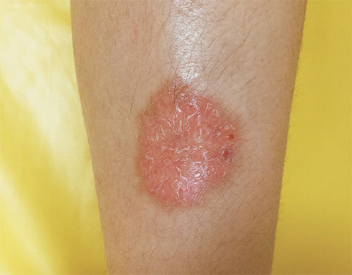
a day with poorly controlled blood sugars. On examination, the girl had
a well-defined, circular, indurated red plaque, measuring 5×5 cm, over
the left leg (Fig.1). The lesion started as painless,
reddish papules that slowly enlarged to a plaque over a period of 3
years. Analysis of the biopsy specimen confirmed the diagnosis of
necrobiosis lipoidica (NL) diabeticorum. Laboratory investigations
revealed an elevated glycosylated hemoglobin (12.5%), normal thyroid
function, normal complete blood count, unremarkable liver and renal
functions, and normal serum cholesterol and triglycerides. She was able
to achieve good glucose control and resume her normal life; however, the
complication on skin persisted despite an intensive insulin treatment
and topical steroids.
 |
|
Fig. 1 Necrobiosis lipoidica plaque
with erythematous margins in the pretibial area.
|
NL is an extremely rare finding in childhood diabetes
and typically presents at 30-40 years of age. The most commonly affected
site is the leg; 85% of cases affect that site exclusively. Differential
diagnoses include granuloma annulare (typically found on the dorsa of
hands, fingers and feet), sarcoidosis, necrobiotic xanthogranuloma,
lichen sclerosus, and erythema induratum. First-line therapy for NL
includes non-steroidal inflammatory agents, cryotherapy and potent
topical glucocorticoid agents for early lesions, and intralesional
corticosteroids injected into the active borders of established lesions.
Systemic glucocorticoid therapy may also be effective, but can be
associated with adverse effects in patients with diabetes.


