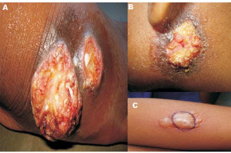
An underweight, 8-year-old boy presented with painless ulcer
on the left retro-auricular region with an undermined edge,
pale granulation tissue on the floor and surrounding bluish
hue. Two painless ulcers were also present on dorsum of
right foot (Fig. 1). Initially the lesions
were firm, erythematous, non-tender nodules which softened
to become abscess and broke down to form ulcers over two
months. Systemic examination revealed no abnormalities.
Investigations revealed anemia (hemoglobin 7 g/dL), high ESR
(60 mm), and a positive mantoux test (20mm).VDRL and ELISA
for HIV were not contributory. The X-ray chest and of
local part were normal. Aspiration cytology from the ulcer
was negative for acid fast bacilli, fungal elements and
anerobic organisms. Histopathology of the lesion showed
granuloma with widespread caseation necrosis. Anti-
tubercular therapy with four drugs regimen was started and
he had remarkable improvement with healing of ulcers and
weight-gain.
 |
|
Fig.1 Multiple deep
seated ulcers with undermined edge on the dorsum of
the right foot (A), and left retro auricular area
(B); Highly reactive mantoux test(C).
|
Tubercular gumma is a rare (1-2%) form of
cutaneous tuberculosis caused by hematogenous dissemination.
Presence of tubercles with widespread caseation necrosis is
diagnostic on histopathology. Clinically, it is difficult to
differentiate from syphilitic gumma, pyoderma gangrenosum,
atypical mycobacterial lession and subcutaneous fungal
infections. It should be confirmed by histopathology and
culture. Pyoderma gangrenosum has a rapidly progressive
course with a invariably painful ulcer. Pathergy phenomena
is positive and histopathology shows neutrophilic
inflammation and necrosis. There is a tendency for central
necrosis and ulceration with peripheral healing and tissue
paper scrapping in syphilitic gumma. Subcutaneous mycoses is
diagnosed by direct microscopic examination with KOH,
histopathology with special stains (PAS, Gomori, Grocott)
and fungal culture.
In this part of the subcontinent the
tubercular gumma should be the first clinical differential
diagnosis with this type of presentation.


