|
A 4-day-old male baby, born after uneventful gestation and vaginal
delivery, was brought for evaluation of cutaneous lesions. The parents had
observed blotchy redness over the face few hours after birth. On
examination, the baby was comfortable, afebrile with no lymphadenopathy.
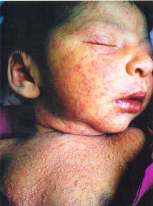
Cutaneous lesions comprised numerous, discrete, blotchy erythematous
macules with a central vesicle or pustule involving face, trunk and limbs,
distributed predominately over chest and proximal extremities (Fig.
1). Except for neonatal jaundice his systemic examination and
investigative profile were essentially normal. Wright’s stained smears
from a vesiclulo-pustule showed eosinophils predominately. A clinical
diagnosis of erythema toxicum neonatorum was made and the parents were
counseled about benign and self limiting nature of the disease. All
pustular lesions subsided on day 6 of life with urticaria-like erythema or
mild desquamation at places.
 |
|
Fig. 1 Small, multiple, erythematous
macules, blotchy at places and topped with central vesicles or
pustules. |
Erythema toxicum neonatorum or toxic erythema of the
newborn is an uncommon, self-limiting, benign dermatosis of unknown
etiology affecting both sexes equally. Its exact prevalence is unclear.
Approxi-mately 50% of full-term infants of all races will manifest some
degree of toxic erythema in first few days of life but it is less common
in premature and small-for-dates babies. The diagnosis is primarily
clinical from typical skin lesions and their characterstics distribution
being more profuse over front of trunk, proximal extremities with
palmoplantar sparing, and face than other body areas. The lesions begin
from birth to tenth day of life as blotchy erythematous macules. They fade
away with in a day in mild cases or evolve into urticarial papules topped
by small pustules within the erythe-matous areas in about 10% cases who
manifest severely, individual lesions clear spontaneously in about 5 days
and by 2 weeks of age all lesions resolve without residual
hyperpigmentation.
These lesions need to be differentiated from other
neonatal pustular dermatoses particularly miliaria rubra or pustular
miliaria, transient neonatal pustular melanosis, bacterial, candida or
Malassezia furfur pustulosis, and most importantly neonatal herpes
simplex infection. Erythema toxicum neonatorum has larger 2-3 cm
erythematous macules as compared to 2-3 cm erythema of miliaria lesions.
Unlike erythema toxicum neonatorum, transient neonatal pustular melanosis
lesions show predominance of neutrophils rather than eosinophils and
resolve with residual pigmentation. Herpes lesions are usually painful,
will coalesce and show multinucleated giant cells in Tzanck smears.
Negative culture for bacteria or fungus and KOH mounts from skin lesions
will exclude neonatal bacterial/fungal infections.
|

