1.gif (1892 bytes)                      

Images

Indian Pediatrics 2008; 45:784

Calcinosis in Juvenile Dermatomyositis


A 5 year old boy was diagnosed to have JDM (Juvenile dermatomyositis) at the age of 2 years and had received treatment with corticosteroids, methotrexate and hydroxychloroquine for three years. The muscle power improved while on therapy. He presented with swelling around the knees and ankles for last two months. X-ray showed massive calcinosis around ankle joint (Fig 1, 2).

Fig. 1 Calcinosis around ankle joint. Fig. 2 Calcinosis around ankle joint (lateral X-ray).

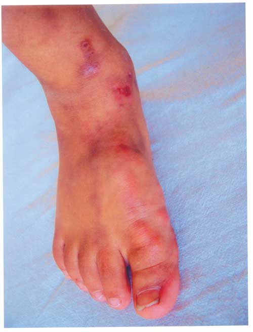
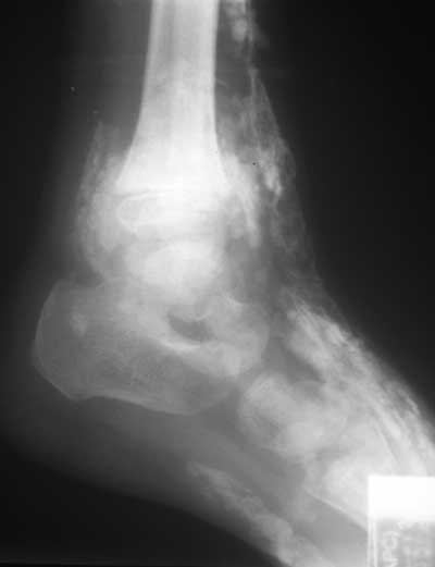
Calcinosis is a sequel of juvenile dermatomyositis. Its pathogenesis is not well understood. It is said to occur in up to 30% of patients with JDM. The sites most frequently affected are the elbows, knees, digits and extremities, although it may occur virtually anywhere over the body. Longstanding active disease, especially when associated with delay in initiation of therapy is supposed to be an important risk factor for development of calcinosis. There is no specific treatment.

Kana Ram Jat,
Surjit Singh,

Advanced Pediatric Center,
PGIMER, Chandigarh 160 012, India:
E-mail: [email protected]


Home

Past Issue

About IP

About IAP

Feedback

Links

 Author Info.

  Subscription