|
|
Images in Clinical Practice Indian Pediatrics 2004; 41:955-956 |
|||||
|
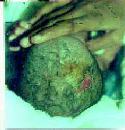
Aplasia Cutis Congenita |
|||||
|
Aplasia cutis congenita is a rare disorder associated with a complete or partial absence of an area of skin. Although this may occur anywhere on the body, about 80% of these lesions involve the scalp, usually the vertex. The size of the lesion may vary from a barely perceptible lesion to lesions larger than 10 cm. The smaller lesions are usually partial thickness defects whereas larger defects are likely to be full-thickness with underlying bony defects in 15-30%. They are known to be associated with cardiovascular, neuro-logical, spinal or chromosomal abnormalities and may be familial. The treatment of scalp defect is aimed primarily at prevention of infection and hemorrhage. An infected lesion can rapidly lead to meningitis. Major hemorrhage especially from the superior sagittal sinus is an ever present risk and is usually preceded by warning bleeds. Small defects are most often of partial thickness and observation or wet dressing (to prevent drying or dessication) is an acceptable option in the management of partial thickness defects, as in our case. The treatment of full-thickness lesion with exposed brain is primarily surgical and is usually managed by full-thickness rotation flaps or split skin grafting. Arvind Sinha,
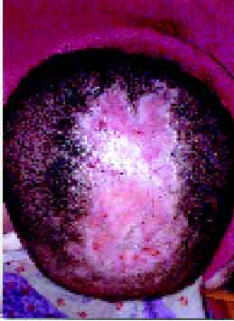
Figure 2 depicts a large area of aplasia cutis on the scalp measuring 10 × 4 × 0.5 cm in another male infant with similar clinical details as the one reported above.
Courtsey: Dr. P.S. Kashyape,
|
![]()