Urological anomalies are common in children born
with anorectal malformations, the incidence and severity being more in
children with high lesions. We report a rare and uncommon case of
posterior urethral valves in a neonate with high anorectal
malformation and solitary kidney.
A 2.2 kg, full term male neonate was referred with
the diagnosis of imperforate anus. Antenatal ultrasound showed
oligo-hydroamnios, but no other malformations. Postnatal history about
urinary stream was not available. On examination, there was absent
anal opening and generalized abdominal distension with no evidence of
meconium tract in the perineum. Invertogram suggested high variety of
anorectal malformation with normal sacrum and vertebrae. The hemato-logical
and biochemical investigations were normal. Preoperative ultrasound
showed dilated bowel loops and a solitary right kidney. 2D Echo was
normal.
The child had a loop sigmoid colostomy formed. In
the postoperative period, he passed urine in drops, and developed
signs of bladder outflow obstruction. A repeat ultrasound showed
dilated bladder with hydroureter and hydronephrosis. A voiding
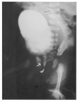
cystourethrogram (VCUG) showed trabeculated bladder and dilated
posterior urethra suggestive of posterior urethral valves with no
evidence of vesicoureteric reflux or any fistulous connection with the
rectum (Fig. 1). A vesicostomy was done on the second
postoperative day, as the smallest available cystoscope was unable to
pass through the child’s urethra. Postoperative recovery was
uneventful. Distal loopogram done 10 days later showed a recto bulbar
urethral fistula (Fig. 2). The child is presently well and on
regular follow up.
 |
 |
|
Fig.1.
Micturating Cystourethrogram suggestive of posterior urethral
valve. |
Fig. 2.
Distal loopogram showing a blind ending rectum with rectobulbar
uretheral fistula. |
Most children with anorectal malforma-tions have
one or more abnormalities that affect other systems. Up to 26-50%
associated urological anomalies have been reported with anorectal
malformations common being renal agenesis, hydronephrosis and
hydroureter (both obstructive and refluxing)(1-3).
The present child had posterior urethral valves
that were diagnosed on the day after colostomy had been performed.
Many authors recommend a renal and bladder ultrasound along with a
renal voiding cystourethrogram in the early neonatal period prior to
colonic diversion(4). However, in view of the abdominal distension,
and preoperative ultra-sound reports, we preferred to decompress the
bowel at the first instance. Postoperative failure of adequate bladder
emptying warranted an early VCUG, which revealed this rare
association.
Evaluation of the most commonly affected organ
systems in patients with anorectal malformations is essential because
it is these associated anomalies that account for most of the
morbidity and mortality(5). The critical combination of bladder outlet
obstruction due to posterior urethral valves, urinary tract infection
due to intestinal fistula and solitary kidney posed a significant
threat to this child.
The severity of the urinary tract anomaly may cause
a high mortality and may preclude useful treatment of the anorectal
malformation. An early thorough attention to these potential sources
of long term morbidity and mortality will ultimately improve survival
and overall quality of life for children with anorectal malformations.
Amar A. Shah,
Anirudh V. Shah,
"Anicare" 13, Shantisadan Society,
Nr. Parimal Garden, Nr. Doctor House
Ellisbridge, Ahmedabad-380 006, India.
E-mail: anirudhshah@icenet.net
