|
|
Case Reports Indian Pediatrics 2003; 40:897-900 |
|
|
Preoperative Aspiration for Anterior Mediastinal Cyst with Respiratory Distress |
|
|
Prema Menon K.L.N. Rao Amita Trehan
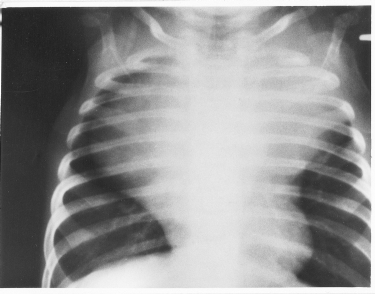
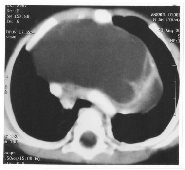
Large mediastinal cysts can present with severe respiratory distress in infancy. Percutaneous guided aspiration is beneficial in such cases to stabilize the patient before surgical intervention. We report the benefits of such an intervention in an infant with an anterior mediastinal cyst. Case Report A 5-month-old boy was admitted with complaints of breathlessness and dry cough of 2.5 months duration, with recent exacerbation of symptoms. There was no history of fever or hemoptysis. There was one episode of cyanosis. There was history of vomiting and difficulty in feeding. On examination, there was pallor, tachypnea, suprasternal and bilateral subcostal recession. Trachea was in the midline. Air entry was equal on both sides. The plain X-ray chest showed a large soft tissue shadow occupying almost the entire upper one-third of the thorax (Fig. 1). The hematological and biochemical parameters were within normal limits. Computed tomography (CT) of the chest showed a cystic lesion occupying the anterior and superior mediastinum (Fig. 2). In view of the respiratory distress and the cystic nature of the swelling, aspiration was performed through the 2nd intercostal space on both sides. About 60 mL of clear fluid was easily withdrawn. This dramatically reduced the symptoms of the patient. A repeat chest X-ray showed a marked decrease in the size of the swelling. Biochemical analysis of the fluid showed a protein content of 20 mg%, glucose of 59 mg% and adenosine deaminase (ADA) of 9 IU/L. Fluid cytology demonstrated clusters of benign columnar epithelial cells, mature squamous cells, lymphocytes and plasma cells. With his condition now improved, the child underwent a left anterolateral thoracotomy.
Fig. I. PA view of the chest radiograph showing a large
anterior
Fig. 2. CT Scan of the chest showing a large anterior
mediastinal The cyst was aspirated again under vision. This not only improved his ventilation but also helped in easy excision of the cyst, which was extending to the opposite side of the chest. Major blood vessels, airways as well as the left phrenic and recurrent laryngeal nerve were identified and protected during surgery. He had an uneventful post-operative course and at 5-month follow-up is asymptomatic. The histopathological report showed an immature teratoma. Discussion Approximately 8% of all mediastinal tumors are benign teratomas, the majority being located anteriorly(1). The tumors are usually partially cystic(2). The primary cystic and neoplastic lesions of the anterosuperior mediastinum include thymomas (30%), lymphomas (20%), germ cell tumors (18%) and carcinomas (13%)(3). Patients with benign teratomas are usually young adults and about 36% are asymptomatic(2). Symptoms pertaining to airway obstruction in mediastinal masses may be present pre- operatively or may develop during induction of anesthesia. The latter may be sudden and life, threatening(4,5). The effect of anesthesia on pulmonary mechanics, the supine body position and the elimination of glottic regulation of airflow by endotracheal intuba-tion have all been postulated as causes of exacerbation of compression of major airways during induction(4). Transcarinal aspiration of a mediastinal cyst following marked inter-ference with gas exchange after institution of general anesthetic has been reported in this context(6). The therapeutic use of transbronchial needle aspiration for a bronchogenic cyst has been reported, although, there was exudative effusion after this procedure(7). Percutaneous aspiration of pericardial cysts without further recurrence has been reported(8). Ethanol sclerosis has also been used in the management of pericardial and thymic cysts(9). Mediastinal cyst aspiration has been attempted during thoracoscopic resection and was found to avoid spillage as well as allow better manipulation and easier grasping of the cyst(10). This benefit was noted in our patient also, wherein the entire lesion including the part extending to the opposite side could be excised easily through a left thoracotomy approach alone. Following aspiration, cyst fluid can rapidly re-accumulate. Aspiration cytology alone does not always give the exact pathology of the lesion and a malignancy may be missed. Therefore, aspiration as the only modality of treatment is not recommended. Complete surgical excision continues to be the treatment of choice. There is no previous case report of percutaneous guided aspiration for alleviating symptoms of respiratory tract obstruction in cystic lesions of the anterosuperior media-stinum in young children. Perioperative symptoms and complications were reduced by aspiration prior to surgery in our patient. We therefore recommend preoperative guided aspiration in large symptomatic cystic lesions of the mediastinum to alleviate patient symptoms and reduce anesthetic risks. Acknowledgement Authors thank Prof. A. Rajwanshi and Prof. R.K. Marwaha for their active role in patient management. Contributors: PM performed drafting of manuscript, literature search and contributed to patient management. AT investigated the patient. KLNR finally approved the manuscript and will act as guarantor for the manuscript. Funding: None. Competing interest : None stated.
|
![]()