Limb body wall complex (LBWC) is a constellation of
severe body-wall defects with evisceration of the organs caused by the
failure to form a body stalk. It is characterized by absent or short
umbilical cord, failure of fusion of amnion and chorion, with associated
severe spinal scoliosis, and limb defects(1-2). We report here a rare
case of this multiple congenital malformation complex with some unusual
associations such as an association with use of oral contraceptive pills
(OCP) by mother during the first 2 months post conception and presence
of an accessory spleen and exstrophy of bladder. Absent thymus and
interstitial calcification in kidney, seen in our proposita, to the best
of our knowledge have not been described earlier.
Case Report
A 22-year-old, unbooked primigravida with severe
oligohydroamnios delivered spontaneously, a 32-week stillborn weighing
1680 grams (including the placenta) at Kasturba Hospital, M.G.I.M.S.,
Sevagram. The proposita had complex fetoplacental malformation pattern
with multiple congenital anomalies. Mother conceived while taking OCP
that she continued to take in the first 2 months post conception. Mother
was not able to recall the exact name of this OCP. There was no history
of consanguinity or any family history of malformations.
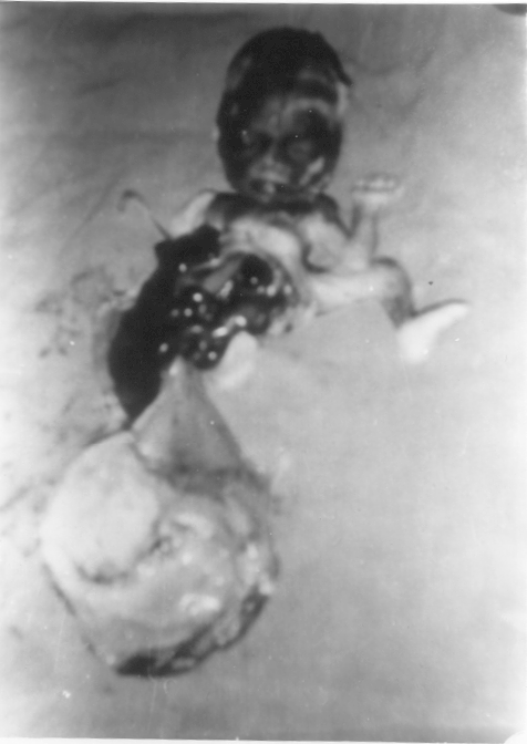
On clinical examination (Fig. 1), the most
obvious features included a huge left-sided abdominal wall defect that
caused evisceration of liver, stomach and the intestines. Abdominal
viscera were attached directly to the placenta and were covered by
ruptured sac of amnion. A rather short and broad body stalk connected
the fetus to the placenta. The umbilical vessels, one artery and a vein,
were embedded in an amniotic sheet, which was connecting the skin margin
of the anterior body wall defect to a circumvellate placenta. The anus
was not visible and no discernable external genitalia were noted. Other
abnormal features included asymmetrical face with medial epicanthal
folds, low set misshapen and elongated ears, compressed nose, high
arched palate and receded chin (Potter facies). Thorax was small and
bell shaped with scoliosis of the spine. The lower limbs had developed
unequally and were malpositioned. Right leg was more hypoplastic with
clubfoot and flexion contracture at the knee joint. The left leg showed
a pescavus deformity.
 |
|
Fig. 1. Newborn with abdominal wall defect,
evisceration of the abdominal organs and lower limb defects. |
Skeletal survey revealed pneumothorax and collapsed
lungs on both the sides. Mandible, maxilla and the nasal bone were
hypoplastic. Upper limbs were normal. There was scoliosis and torsion of
the spine. The long bones of the right lower limb were underdeveloped
with hypoplastic clubfoot. There was splaying of pelvic bones and
hypoplasia of the sacrum. The cranial ultrasound examination (USG) was
normal.
Autopsy confirmed the above skeletal and USG
abnormalities. A huge abdominal wall defect, extending from the distal
part of the sternum to the suprapubic region, involving more of the left
side was observed. This caused evisceration of liver, stomach and the
intestines. Margins of liver were ill-defined and scattered over the
ruptured membranes of amnion. An accessory spleen was present in
addition to a normal spleen. The colon ended blindly in a large meconium
filled pouch; there was anal atresia. The urogenital anomalies included
hypoplastic kidneys and absent gonads. The ureters drained into a
rudimentary bladder, which opened to the exterior. Thymus was absent and
both the lungs were hypoplastic. Diaphragm was present. Heart showed a
large atrial septal defect and primitive single ventricle.
Histopathology of the kidneys, and the lungs showed marked immaturity.
Right kidney showed interstitial calcification. Liver showed
extramedullary haemopoiesis. Gonads could not be localized but a mass of
suspected gonad, which was embedded in the membranes, was histologically
detected as testicular tissue. Karyotyping was not possible due to
culture failure.
Discussion
Limb body wall complex (LBWC) represents a set of
disruptive abnormalities having in common the failure of closure of the
ventral wall, and a shortened or absent umbilical cord(1,2) There is
failure of fusion of the amnion and chorion. The diagnosis of this
entity is based on two of the three following characteristics (a)
exencephaly/ encephalocele and facial clefts; (b) thoraco and/or
abdominoschisis; and (c) limb defects(2-5). The association of
any abdominal wall defects and scoliosis implies the diagnosis of
LBWC(3). ln our proposita, the two diagnostic features were
abdominoschisis and limb defects, further supported by severe torsion
and scoliosis of the spine.
LBWC is also referred to as "congenital absence of
the umbilical cord", "cyllosomus and pleurosomus" and "body stalk
anomaly"(1-3) The incidence of LBWC is 1 : 14,273 birth(6). There is no
sex predilection and the risk of recurrence is negligible(1,3). The
etiology of this anomaly is not clear. Chromosome defects have not been
identified. In a largest collection of 25 fetuses with LBWC reported by
Van Allen, et al.(2), pregnancy abnormalities by history included
removal of intrauterine device 6 weeks post last menstrual period in one
mother; tubal ligation with general anesthesia 4 weeks post conception
in one; reported illicit drug abuse in two; and use of birth control
pills during the first two months post conception in one. Till date no
other case of LBWC in association with birth control pills during the
first trimester of pregnancy is reported. There is a strong association
with cocaine abuse during the first trimester of pregnancy(7). ln our
case there was history of exposure to birth control pills (exact
preparation not known) during the first 2 months post conception.
However more observations are required to comment on the role of birth
control pills in the etiopathogenesis of LBWC.
The pathogenesis of limb body wall complex is
uncertain though two major hypotheses are currently formulated. One
theory is an early rupture of the amnion with mechanical compression
between the 3-5th embryonic weeks(1,3). The other theory is the vascular
disruption theory(1,2). Malformation of the body stalk results from a
defect in the germ disc leading to an abnormal body folding, an abnormal
amniotic cavity formation and a failure to obliterate the extraembryonic
coelom. This accounts for absent or short umbilical cord and broad
insertion of the amnio-peritoneal membrane onto the placental chorionic
plate(3). Aplasia or hypoplasia of the paraspinous or thoracolumbar
musculature is responsible for the severe scoliosis(2,6). The limb
defects are due to the mechanical rupture through the amnion in the
presence of a persistent extraembryonic coelom(3,6).
Limb defects in LBWC are seen in 96% cases and
include club foot (32%), oligodactyly (12%), arthrogryposis/web (12%),
absent limb (9%), single forearm bone (8%), single lower leg bone (6%),
pseudosyndactyly (5%), split hand/foot (5%), radial/ulnar hypoplasia
(4%), rotational defect (4%), and preaxial poly-dactyly (3%)(5).
Internal malformations in LBWC are seen in 95% cases
and include cardiac anomalies (43%), absent diaphragm (74%), abnormal
pulmonary lobulations (50%), gastrointestinal (100%), trilobulated liver
(4%), polysplenia (4%), absent gall bladder (29%), renal (65%) and
urogenital abnormalities (56%). Other malformations include amniotic
bands (40%) and single umbilical artery(1-3,8). Cardiac malformations
reported are primitive ventricle (53%), common atrium (46%), truncus
arteriosus (23%), atrial septal defect (15%), membranous VSD (8%),
hypoplastic right ventricle (8%) and ectopia cordis (8%).
Gastrointestinal anomalies seen are nonrotated intestine (96%),
intestinal atresia (22%), anal atresia (17%), shorted intestines (4%)
and Ladd’s bands (4%). Renal abnormalities reported are unilateral
absent kidney (30%), bilateral absent kidney (4%), hydronephrosis (17%),
renal dysplasia (9%), and hypoplastic kidneys (4%). Urogenital
abnormalities seen are abnormal external genitalia (32%), absent gonad
(30%) and extrophy of bladder (4%) (2). Absent thymus and interstitial
calcification in kidney, seen in our proposita, have not been described
earlier.
In view of the dismal prognosis, early antenatal
diagnosis is important allowing for earlier, and less traumatic
termination of pregnancy(3). Prenatal diagnosis is possible by detection
of very high maternal serum alpha-fetoproteins and by transvaginal
ultrasound examination at the end of the first gestational
trimester(1,6). Postnatally, the examination of placenta, umbilical cord
and the membranes is crucial in confirming the diagnosis of LBWC(3,4).
Contributors: SM diagnosed the condition and
drafted the manuscript; he will act as guarantor. NG performed autopsy
and histology. PC and KYV co-drafted and critically evaluated the
manuscript.
Funding: None.
Competing interest: None stated.
