A 6-year-old boy presented with complaints of
hoarseness of voice since infancy and multiple scarred lesions over face
for past three years. There was a preceding history of recurrent mildly
pruritic papulo-vesicular eruptions over face and distal extremities.
The child was born of non-consanguineous marriage and no other family
members were affected. There were no history of seizures or
photosensitivity, and developmental milestones were normal.
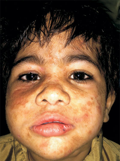
On physical examination, multiple pock-like atrophic
scarring along with few waxy papules were seen on face (Fig.
1) and dorsum of both hands. Lower lip was enlarged and protuberant.
The margins of both upper and lower eyelids were beaded with yellowish
waxy papules (moniliform blepharasis or string of beads). Loss of
eyelashes was also apparent on inner side of right lower eyelid.
Histopathologic examination of skin lesions revealed deposition of
periodic acid-Schiff (PAS)-positive eosinophilic hyaline materials
within the dermis along with adnexal atrophy and onion skinning of blood
vessels. All these findings were suggestive of Urbach-Weithe disease.
 |
|
Fig. 1 Pock-like atrophic scarring and
waxy yellowish papules on face, along with lower lip, thickened
lower lip, beaded eyelid margins with yellowish waxy papules and
loss of eyelashes on inner side of right lower eyelid.
|
Urbach-Weithe disease, also known as lipoid
proteinosis, is an uncommon autosomal recessive disorder caused by loss
of function mutation in extracellular matrix protein-1 gene
leading to aberrant deposition of hyaline materials (lipid-protein
complexes) within skin, oral mucosa, vocal cords and occasionally
intraocular structures. The differential diagnoses are erythropoietic
protoporphyria, amyloidosis and papular mucinoses. Absence of
photosensitivity, urine and stool tests for porphyrins and absence of
mucin and amyloid deposition in tissue samples rule out these entities.
There is no definitive cure of this condition. Oral
dimethylsulphoxide, penicillamine and etretinate have been used with
variable efficacy. Laryngeal lesions can be treated with
microlaryngoscopy and dissection of the vocal cords. Dermabrasion,
chemical peeling and laser therapy is helpful for skin lesions.


