|
|
|
Indian Pediatr 2016;53: 936 |
 |
Aspiration of Rear End of Pen in Children –
Management Issues
|
|
S Kalpana And B Sarath Balaji
Department of Pulmonology, Institute of Child Health
and Hospital for Children, Egmore, Chennai 600 008, India.
Email: [email protected]
|
|
Rear pen-ends are unusual foreign bodies in the airway, occurring mostly
in school-age children. A total of seven children (age 3-9 y), all boys,
were admitted in our hospital with history of aspiration of detachable
pen end from January 2012 to December 2015. The duration from the
aspiration to presentation was from 10 days to as late as 2 months. Only
one child presented acutely as the pen-end had lodged in the subglottis.
The most common presenting complaint was cough (7/7), followed by noisy
breathing in the form of audible wheeze (3/7), and fever and breathing
difficulty (1/7 each). Most of them (6/7) had received repeated
nebulizations prior to their presentation. Air-entry was equal in five
children; 2 children had reduced air-entry on the side of the
aspiration. A monophonic wheeze was the only added sound heard (5/7);
one child had stridor. Chest X-ray was normal (3/7) or showed
unilateral hyper-inflation (3/7) or pneumonic consolidation (1/7).
During flexible bronchoscopy, the pen-ends were seen on left side in
three, right side in three, and subglottis in one child. The sites of
lodgement were in the main bronchus (3/7), segmental bronchus (2/7),
subsegmental bronchus (1/7) and subglottis (1/7). Successful removal by
rigid bronchoscopy was possible in four children. The rest underwent
segmental lobectomies after multiple failed attempts at rigid
bronchoscopic removals. All seven children recovered uneventfully after
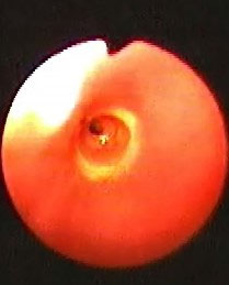
removal with no post-removal or post-surgical complications. All the pen
caps that were ultimately retrieved, either by rigid bronchoscopy or by
surgery, had a perforation at the rear end (Fig.1). This
hole, which had allowed sufficient air entry when lodged in the
bronchus, explains the absence of breathing difficulty or respiratory
distress as a presenting feature [1].
 |
|
Fig. 1 Flexible bronchoscopic view
showing rear end of pen in a segmental bronchus with overlying
granulation tissue and central hole.
|
Tapering rear end of most of the pen-ends causes them
to be lodged in the segmental or subsegmental bronchial segments. The
mouth of the pen cap is always upwards and moves through the glottis
into the airway, and easily forms an inlay with the bronchus, making
removal a big challenge [2,3]. In all the three cases which required
lobectomies, the pen-ends were lodged in the segmental and subsegmental
bronchi, covered by granulation tissue and were simply not visible with
the rigid bronchoscope.
Aspiration of a perforated pen-end neither presents
with the classic clinical features like respiratory distress and
decreased breath sounds nor the typical secondary radiographic changes.
Therefore, high clinical suspicion and use of flexible bronchoscopy as
the initial technique of evaluation in patients with suspected foreign
body aspiration are needed. Given the high probability of aspiration,
pens without detachable rear ends should be encouraged for use in
children.
Contributors: KS, SBB: case management,
conception of the study, acquisition of data and drafting the
manuscript. All the authors approved the final version of manuscript.
Funding: None; Competing interests:
None stated.
References
1. Seifarth FG, Triana J, Knight CG. Aspiration of a
perforated pen cap: complete tracheal obstruction without radiologic
evidence. Ann PediatrSurg. 2012;8:22-4.
2. Can D, Yilmaz O, Astroy S, Gulle S, Yukrel H.
Aspiration of foreign bodies that allow air passage through. Open J
Pediatr. 2011;1:90-93.
3. Wu J, Gu M, Wang Z, Li X. A clinical analysis of
21 cases of pen sheath bronchial foreign bodies in children. Int J Clin
Exp Med. 2015;8:1108-14.
|
|
|
 |
|
