|
|
|
Indian Pediatr 2014;51: 849 |
 |
Atypical Herpes Zoster
|
Bijay K Meher, Deepti D Pradhan and Subhasmita
Pattanayak
Department of Pediatrics, Sardar Vallavbhai Patel
Post Graduate Institute of Pediatrics, SCB Medical College,
Cuttack, Orissa, India.
Email: [email protected] n
|
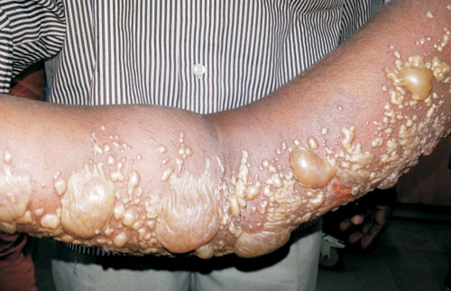
A 12-year-old girl – who was a diagnosed case of connective
tissue disorder and was on oral prednisolone (2 mg/Kg/d) and
hydroxychloroquine for 6 months – presented with multiple
vesiculobullous eruptions over the right hand for 4 days.
Eruptions started with tingling sensation and pain. On
examination, she had steroid facies, hypertrichosis and
hirsutism. Vesicobullous lesions were present on the
dermatomal involvement of C8 and T1 (Fig. 1).
Investigations revealed normal blood counts erythrocyte
sedimentation rate, C-reactive protein, liver function tests
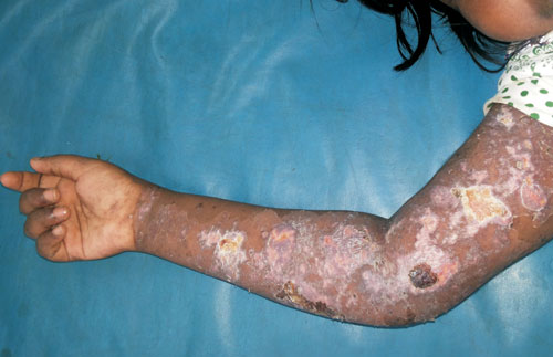
and renal function tests. Tzanck smear revealed
multinucleated giant cells. A diagnosis of herpes zoster
infection was made and patient was started on intravenous
acyclovir (10mg/kg every 8 hrly); skin lesions healed within
7 days (Fig. 2).
 |
|
Fig. 1 Vesiculobullous
lesions over C8 and T1 dermatone.
|
 |
|
Fig. 2 Lesions showing
healing after 1 week of intravenous acyclovir.
|
The diagnosis of herpes zoster is usually
based on unilateral pain in a defined area accompanied by a
typical rash in the dermatomal distribution of a segmental
nerve.
Manifestations of herpes zoster in immunocompromised
children can be severe and life threatening. Patients with
high risk for disseminated disease should receive
intravenous acyclovir at 10 mg/kg every 8 hrly. Patients
with uncomplicated herpes zoster and low risk for visceral
dissemination should be treated with oral acyclovir,
famciclovir or valacyclovir.
|
|
|
 |
|
