A full term newborn boy, weighing 3 kg,
born out of an uncomplicated pregnancy, was brought to us
for evaluation of a few small, white and round bumps on the
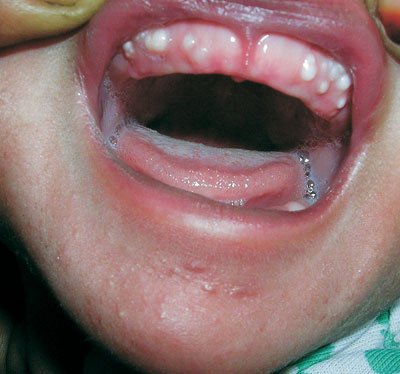
gingival surface. Examination of the oral cavity showed
multiple, firm, pearly-white papules measuring 2 to 4 mm in
diameter, grouped over the vestibular aspect of the alveolar
ridge of the maxillary arch (Fig. 1). Two
similar lesions were seen on the mandibular area. These
lesions were asymptomatic, non-tender, and fixed to the
mucosa. Oral mucosa was otherwise normal. A few milia were
noted on his chin. Detailed systemic examination was normal.
No specific therapy was prescribed. Within a couple of
months, most of the lesions subsided spontaneously. Based on
the clinical features and the natural course of the disease,
a diagnosis of Bohnís nodule was made.
 |
|
Fig. 1 Pearly-white papules
(Bohnís nodules) on alveolar ridge of a neonate.
|
Bohnís nodules are keratin cysts derived
from remnants of odontogenic epithelium over the dental
lamina or may be remnants of minor salivary glands. They
occur on the alveolar ridge, more commonly on the maxillary
than mandibular. Common differential diagnoses include other
developmental oral inclusion cysts (Epstein pearl, Dental
laminar cyst) and natal teeth. Epstein pearl is a small,
firm, white, keratin-filled cyst, located on the mid
palatine raphe. Dental laminar cyst (gingival cyst) is a
yellow-white cystic lesion over the alveolar crest that
arises from epithelial remnants of the degenerating dental
lamina. Natal teeth usually erupt in the centre of
mandibular ridge as central incisors. They have little root
structure and are attached to the end of the gum by soft
tissue.
Bohnís nodules usually rupture
spontaneously and disappear within a few weeks to a few
months. Counseling of the family members regarding its
benign and self-limiting nature is all that is required in
the management.

