A 14-year-old female presented with reticulate pigmentary
changes since the age of 4 years. Initially, the lesions
were present only over the fingers and gradually they
increased in size as well as number to attain the present
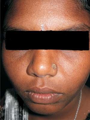
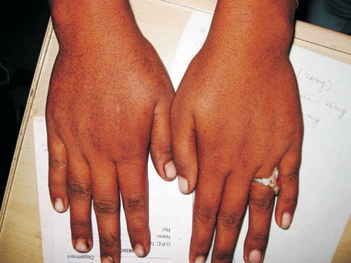
status. Cutaneous examination revealed sharply demarcated
atrophic hyperpigmented macules over the dorsum of
extremities and upper as well as lower eyelids of both
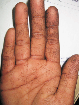
sides. Another important finding was presence of palmar pits
and breakage of epidermal ridge pattern (Fig. 1 to
3). There wasn’t any hypopigmented lesion anywhere.
Chest, abdomen, lower extremities and flexures were spared.
Examination of hair, nail and mucosa did not reveal any
abnormality. Our patient’s mother had similar lesions. Skin
biopsy from a hyperpigmented macule over the dorsum of hand
showed increased melanin in the basal layers. Based on these
findings, a diagnosis of Reticulate acropigmentation of
Kitamura was done. Important clinical differential diagnoses
include dyskeratosis congenita (poikiloderma,
leukoplakia and nail dystrophy), Dowling Degos (reticulated
hyperpigmentation of major flexures, comedones on the back
and neck and pitted facial scars), dyschromatosis symmetrica
hereditaria (symmetrical dyschromatosis of distal
extremities, especially dorsal aspects of hands and feet but
palms, soles and mucous membranes are spared),
dyschromatosis universalis hereditaria (generalized
hyper-and hypopigmented macules; can occur on palms and
soles, but not mucous membranes).
 |
|
Fig. 1 Hyperpigmented
macules on face.
|
 |
|
Fig. 2 Hyperpigmented
macules on dorsum of hands.
|
 |
|
Fig. 3 Palmar pits and
broken epidermal ridge pattern.
|
Reticulate acropigmentation of Kitamura
is characterized by a network of freckle-like areas of
pigmentation which develop on the dorsa of the hands in the
first two decades, and may subsequently involve most parts
of the body. Several individual cases and families have been
reported with features of both Kitamura’s disease and
reticulate pigmented anomaly of the flexures (Dowling–Degos
disease). Mode of inheritance is primarily autosomal
dominant. Such conditions are usually refractory to therapy
and reassuring the patient is the best modality that can be
offered.

