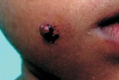
A four-year-old girl presented with an asymptomatic lesion
over the right cheek of two months duration. History of
precedent trauma and bleeding on manipulation was present.
There was no drug intake or systemic complaints. Cutaneous
examination showed a solitary, soft, non-tender, vascular
papule with surrounding hemorrhagic crust over right cheek (Fig.
1). Systemic examination was normal. A clinical
diagnosis of pyogenic granuloma was confirmed by excisional
biopsy.
 |
|
Fig.1 Multiple deep
seated ulcers with undermined edge on the dorsum of
the right foot (A), and left retro auricular area
(B); Highly reactive mantoux test(C).
|
The term pyogenic granuloma (PG) is a
misnomer, as it is neither infectious nor granulomatous. It
is a reactive vascular nodule usually occurring after injury
or irritation. PG is commoner in children and young adults,
with no sexual predilection. PG may be associated with drugs
like systemic retinoids, indinavir, etc.
Clinically, PG presents as a solitary,
painless, red, brownish-red or bluish black papule/nodule.
It is partially compressible without pulsation. Common sites
affected are fingers, feet, lips, head, upper trunk, oral
mucosa and perianal area. Spontaneous disappearance is rare.
Histologically, a well circumscribed proliferation of small
capillaries in a lobular pattern is seen. Differential
diagnoses are hemangiomas, glomus tumors, warts and
molluscum contagiosum. Hemangiomas are scarlet-red,
non-tender, compressible, dome shaped papules/nodules
present at birth/early infancy characterised by early rapid
proliferation and spontaneous involution by 1-9 years of
age. Glomus tumors are multiple, congenital, red/blue, less
compressible, painful/painless papules coalescing to form
plaques over any part of the body. Warts are multiple,
asymptomatic, verrucous papules anywhere over the body; when
inflamed, they may resemble PG. Molluscum contagiosa are
multiple, pearly/skin coloured umbilicated papules; when
infected/irritated they may simulate PG. However, all these
conditions may be differentiated from PG by histopathology.
Complications of PG include ulceration, profuse bleeding,
development of satellite lesions or disseminated, eruptive
occurrence. Treatment includes curettage with cauterization
of the base, surgical excision, sclerotherapy, cryosurgery
and pulsed dye laser.


