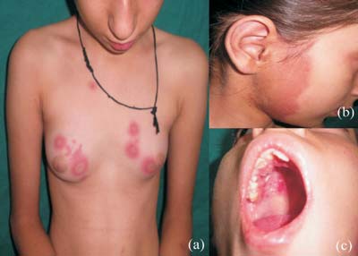
A 13-year-old female child presented with multiple
erythematous papules and plaques with some of them showing
targetoid appearance (Fig.1a), along with a
single large 8 cm x 10 cm sized polycyclic, erythematous
plaque, involving right side of face, upper neck and
external ear area (Fig.1b). Oral mucosa showed
erythematous plaque involving right side hard palate with
overlying multiple superficial erosions (Fig.1c).
She had history of low-grade fever, photosensitivity, malar
rash, Raynaud’s phenomenon, chilblains and recurrent oral
ulceration since 6 months. There was no history suggestive
of recent infection or drug intake prior to onset of
lesions.
 |
|
Fig.1 Rowell syndrome
|
Laboratory studies showed positive
antinuclear antibodies (ANA) in a speckled pattern at 1:160
dilutions, ESR 25 mm/I/h, and positive rheumatoid factor.
dsDNA antibody, AntiRo and antiLa antibodies were negative.
Hemogram, C3, C4, urine routine, urinary 24-hr protein, and
renal function test were all within normal limits.
Electrocardiogram and chest X-ray were normal.
Histopathology of skin lesion was consistent with the
diagnosis of systemic lupus erythematosus (SLE). Patient was
advised photo protection and started on tapering oral
corticosteroid along with Hydroxy-chloroquine. Lesions
resolved after one month of treatment leaving only mild
pigmentary changes with no relapse at 6-month follow up.
Rowell Syndrome (RS) is a unique clinical
association of lupus erythematosus (LE) with erythema
multiforme (EM) like lesions and a characteristic
immunologic pattern. The etiopathogenesis is unknown.
Diagnostic criteria include three major criteria: (1) LE
(systemic, discoid, or subacute); (2) EM-like lesions with
or without mucosal involvement; and (3) speckled ANA; and
three minor criteria: chilblains, antiRo or antiLa
antibodies, and positive rheumatoid factor. For diagnosis
all three major and at least one minor criterion should be
fulfilled. Children with LE may present with EM-like lesions
which must be differentiated from Classical EM which is
often precipitated by factors such as infective agents or
drugs and is not associated with any specific serological
abnormalities or with chilblains. Treatment is with topical
and oral steroids, dapsone, and antimalarial agents but
response is variable with frequent recurrences.


