|
|
|
Indian Pediatr 2011;48:
829 |
 |
Pachyonychia Congenita Affecting only Nails |
|
Piyush Kumar, Panchami Debbarman and Avijit Mondal,
Department of Dermatology, Medical College and Hospital,
88, College Street,
Kolkata 700 073, West Bengal.
Email: [email protected]
|
|
A 9 years old boy presented with painful discolored and thickened nails of
all 20 digits since birth. Antenatal, natal and immediate postnatal
history was normal. At birth, parents noticed discolored nails of all
digits. Over time, they thickened and became deformed, causing occasional
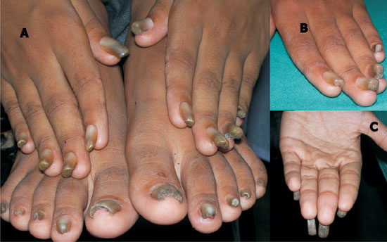
pain and difficulty in holding small objects. On examination, all nails
were discolored and thickened. There was transverse overcurvature,
increasing along the long axis of nail from proximal to distal end. As a
result, distal free end was most curved (Fig.1). Palms,
soles and mucosae did not reveal any abnormality. Rest of the
mucocutaneous examination was noncontributory. Diagnosis of pachyonychia
congenita was made. Pachyonychia congenital affecting only the nails is
extremely rare.
 |
|
Fig. 1 Discolored, thickened, deformed
nails of all digits (A). Close up view- "Omega" shaped distal end of
nail (B). Note absence of palm involvement (C). |
The presentation is quite distinctive and hardly poses
any diagnostic difficulty. Trauma and rarely, onychomycosis may result in
pincer nail. However; it is limited to few nails only. Hereditary pincer
nail syndrome (having distally directed exostoses on the tibial side of
all toe phalanges, an obligatory criterion and may have destructive
arthrosis of the terminal joints of digits) and pachyonychia congenita
have pincer nail deformities of all nails. Pachyonychia congenita is a
rare genodermatosis caused by mutation in keratin gene and is
characterized by dystrophic, thickened nails (pachyonychia), symmetric
focal palmoplantar keratoderma and oral leukokeratosis. Pachyonychia
congenita has been divided mainly into 2 types: PC type 1 (more common,
also known as Jadassohn-Lewandowski syndrome) and PC type 2 (also known as
Jackson-Lawler syndrome). Palmoplantar keratoderma and oral leukokeratosis
are less severe and less frequent in type 2. The distinguishing features
of type 2 are natal teeth, steatocystomas, and pili torti. The treatment
is mainly surgical with the aid of dermal grafts or partial matricectomy.
Conservative treatment with urea 40% or steel brace has met with success;
however, treatment has to be continued for a long time.
|
|
|
 |
|

