| |
|
Correspondence Indian Pediatrics 2008; 45:866-867 |
|||
|
Acute Hemorrhagic Edema of Infancy |
|||
|
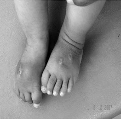
A 7 month-old male was admitted with sudden onset edema of dorsum of hands and feet, multiple spontaneous ecchymotic spots over face and limbs and low-grade fever. Before he presented to us, the child had undergone incision and drainage of the dorsum of the foot on 2 occasions, the fluid drained was sero-sanguinous. Examination revealed edema of hands and feet (Fig.1) and swelling of inter-phalangeal joints. Over the next 2-3 days edema and ecchymosis almost disappeared without any specific therapy. Another 4 days later, he again developed edema of the whole right lower limb and dorsum of left foot, restricted extension of the right knee joint and circinate skin lesions on the face. A clinical diagnosis of AHEI was considered at this stage.
Laboratory investigations showed progressive anemia (hemoglobin 6.3g/dL), rising ESR (up to 60mm/h) with thrombocytosis (13 × 103/µL). Other investigations to rule out mimicking conditions were normal. Skin biopsy from lesion on face showed luminal narrowing and destruction of the elastic lamina of the small and medium sized arteries. Parents were reassured and sent home without specific therapy. After a waxing and waning course over 4-6 weeks, the child improved. AHEI is a distinctive cutaneous, small vessel leucocytoclastic vasculitis. It is common in males without racial predilection, usually benign and without sequel and with spontaneous recovery within 1-3 weeks(2,3). It presents at 4-24 months and occurs during winter(4,5). It is usually preceded by viral infections, drug intake or vaccination(2). Clinical findings develop rapidly over 24-48 hrs. The two primary features include (i) large cockade (rosette or knot of ribbons), annular or targetoid purpuric lesion found primarily on face, ears and extremities, and (ii) non-tender edema of the limbs and face. It may be asymmetric. Low-grade fever is common(2). Recurrent episodes may occur. Arthritis, nephritis, abdominal pain, GI bleeding and lethal intestinal complications are rare(2). The entity should be differentiated from erythema multiforme, HSP, and (iii) meningococcemia(2). A slightly prolonged course of the disease with anemia, thrombocytosis and elevated ESR were unusual features in our patient. Acknowledgment We thank Dr Raju Khubchandani for his advise in management as well as manuscript preparation, Dr Shaila R Khubchandani for histopathological diagnosis, and Dr Rajkumar Dheer for assistance in management of the patient. Gautam Jain,
|
![]()