|
|
Case Reports Indian Pediatrics 2007;44:781-782 |
||
|
Capillaria hepatica Infestation |
||
|
Fazal Nabi From the Department of Pediatrics & Histopathology, Jaslok Hospital & Research Center, 15, Dr. G. Deshmukh Marg, Mumbai 400 026, India. Correspondence to: Dr. H.K. Palaha, Pediatric
Intensivist, Jaslok Hospital & Research Centre, 15, Dr. G Deshmukh
Marg, Mumbai 400 026, India.
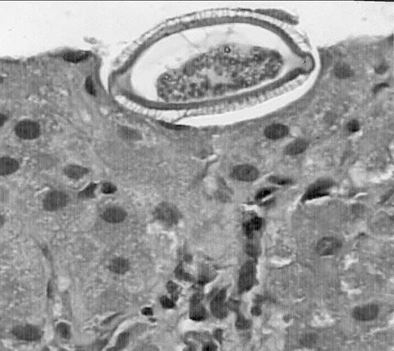
Abstract: Capillaria hepatica primarily infests rodents and is rarely found in humans. Till date only 37 cases have been reported worldwide out of which two are from India. We hereby report a fourteen-month-old child who presented with pyrexia of unknown origin; later diagnosed to have Capillaria hepatica infestation. A fourteen-month-old male child, resident of Nasik, was brought to our hospital with moderate to high grade fever for one month. This was preceded by loose stools for three days. On examination, the child was active and playful and weighed 10 Kg. Systemic examination was normal except for mild hepatomegaly. Investigations revealed hemoglobin 8.7 g/dL, total leucocyte count–24,900/cumm, polymorphs–18%, lymphocytes–25%, eosinophils–55% and monocytes–2%. Liver function tests revealed mildly raised liver enzymes, viz., alanine aminotrans-ferase–116 IU/L, aspartate aminotransferase– 138 IU/L. Ultra-sound of the abdomen revealed mild hepatomegaly with a liver span of 9.7 cm, no intrahepatic biliary canalicular (IHBC) dilatation; portal vein and common bile duct were normal. There were enlarged lymph nodes in porta hepatis region, the largest one measuring 9 mm. CT abdomen revealed retropancreatic and preportal lymph nodes which were subcentrimetric. Bone marrow study was normal. Meanwhile, the child continued to have moderate grade fever and was given symptomatic treatment. A liver biopsy was planned in absence of any diagnostic clues. The histopathological examination revealed classic spindle shaped eggs of nematode Capillaria hepatica. The eggs forms were pathog-nomic with outer striated wall and polar plugs on both end of the spindle. Some fibrosis was also seen (Fig.1). Albendazole was started and child became afebrile, liver size regressed, serum transaminase levels became normal within 2 weeks of starting therapy.
Capillaria hepatica is zoonotic nematode, primarily found in the liver of the rats(1). Human beings are accidental hosts. The unembryonated eggs of Capillaria hepatica are found in the liver of the first host and reach the intestine of the second host when the first host is eaten by a predator, or undergoes natural decomposition after death, whereby these eggs are released in the soil. They become embryonated in the soil and on ingestion by second host by feco-oral contamination, or geophagy, reach the intestine of second host. Here they migrate via the walls of intestines and through the portal vein, reach the liver where they grow into adult forms(2). This is known as genuine infestation and produces symptoms like persistent fever, hepatomegaly and cosinophlia(3). The differential diagnosis includes accidental tissue infestation by nematodes including. Toxocara cati, Toxocara canis, Fasciola hepatica and visceral larva migrans. Liver biopsy remains the cornerstone of diagnosis, but serological testing by indirect immuno-fluorescence assay is also recommended for diagnostic and screening purpose(4). So far in the world literature only 37 cases of this disease have been reported, out of which 16 have been children(4). Two cases have been reported from India so far(5). Pica is an important presenting complaint, and this explains why this parasite infests the younger especially those between 1 and 5 years of age (94%). Anemia is frequently observed and so is hypergammaglobulinemia(3). Treatment of choice is thiabendazole for 3 months. Alternative drugs include albendazole, and possibly ivermectin. Steroids are sometimes used to reduce hepatic inflammation(6). Prognosis depends on severity of infestation. With massive destruction of liver tissue, hepatic failure and death ensue if liver transplant is not available. Contributors: FM Managed the case, HKP prepared the manuscript. DS worked up the case, AC did the histopathological reporting. Funding: None. Competing Interests: None stated. | ||
|
References | ||
|
![]()