|
|
Images in Clinical Practice Indian Pediatrics 2005; 42:1046-1047 |
||
|
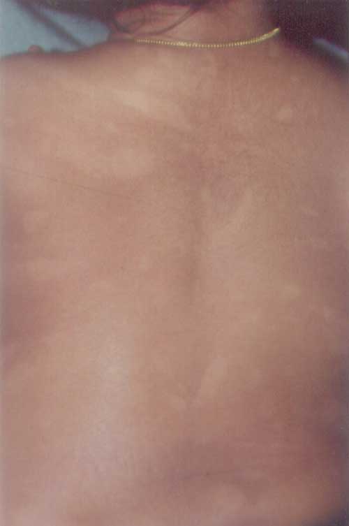
Systematized Nevus Depigmentosus |
||
|
ND is a congenital, non familial, well circumscribed, uniformly hypopigmented macule, stable in its relative size and distribution throughout life and involving predominantly the trunk and proximal extremities and rarely the head and neck. Both sexes are equally affected and there is no distinct pattern of inheritance. Histologically there are normal to decreased number of melanocytes with normal size, shape and melanin content of melanosomes, but reduced number and aggregated within vacuoles. Clinical diagnostic criteria for ND include (i) leukoderma present at birth or onset early in life, (ii) no alteration in distribution of leukoderma throughout life (iii) no alteration in texture, or change of sensation, in the affected area and (iv) no hyperpigmented border around the achromic area. Three morphological variants of ND have been described; isolated (circular or rectangular), which is the commonest presentation, segmental and the rare systematized (unilateral whorls or streaks) variety. Systemic manifestations are rare in ND, although systematized ND in association with seizures, mental retardation, limb hemihypertrophy, atopic dermatitis, yellow scalp hair, has been described. Systematized ND is often confused with hypomelanosis of Ito (incontinentia pigmenti achromians). It presents as unilateral or bilateral hypo-pigmented streaks and whorls that follow Blaschko’s lines, the lesions exhibit changes in their manifestation over time (initially progress and then regress), are associated with ocular, musculoskeletal and CNS abnormalities in 62% to 94% of cases and show familial tendency (autosomal dominant mode of inheritance in some cases). Other differential diagnosis in children who present with hypopigmented macules include vitiligo, piebaldism, nevus anemicus, ash-leaf macules of tuberous sclerosis, pityriasis alba, pityriasis versicolor, hypopigmentation of post kala-azar dermal leishmaniasis (PKDL) and post inflammatory hypopigmentation. Sujay Khandpur,
|
![]()