|
|
Images in Clinical Practice Indian Pediatrics 2004; 41:1060-1061 |
||||
|
Spondylocostal Dysostoses |
||||
|
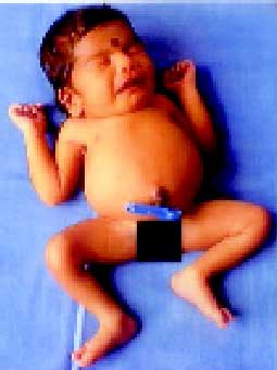
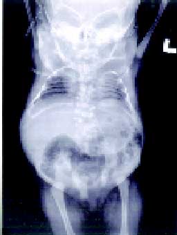
Spondylocostal dysostoses are a heterogeneous group of disorders characterized by multiple vertebral and rib anomalies. Affected individuals have short trunked dwarfism of prenatal onset associated with non-progressive kyphoscoliosis, and restrictive lung disease that is usually the cause of early death. Radiologically, the disease is characterized by vertebral malformations including hemivetebrae, block vertebrae, fused vertebrae and spina bifida and deformities of the ribs that include absent ribs and bifid or fused ribs, which give the typical "crab like", or "fan like" appearance. Associated malformations include those of the urogenital system, cardiovascular system, central nervous system and neural tube defects. Terminology in literature is confusing, as multiple names have been used for similar clinical syndromes. However, patients with multiple vertebral segmentation defects can be classified into three distinct entities based on radiographic and clinical findings: Jarcho-Levin syndrome, a lethal autosomal recessive form, characterized by a symmetric crab-chest and death due to respiratory failure in infancy; spondylocostal dysostosis, a benign autosomal dominant condition; and spondylo-thoracic dysostosis, which shows consider-able clinical and radiographic overlap with spondylocostal dysostosis and has an autosomal recessive mode of inheritance. It has been shown in some autosomal recessive SCD families that the defective locus is on chromosome 19q13. Subsequent mutation analysis has determined that mutations in the human somitogenesis gene, delta-like 3 (DLL 3), which encodes a legend for the Notch signalling pathway causes autosomal recessive SCD. Prenatal ultrasound diagnosis is possible and characteristically shows the presence of fanned out ribs from fused thoracic vertebral bodies. Reconstructive surgery of the chest including titanium rib implants has been described in the treatment of this disorder. Niranjan Thomas, |
![]()