P
erianal abscesses are soft
tissue infections of the perianal region and are common in infants
[1-3]. Most of them are idiopathic, although there may be an association
with congenital abnormalities of the crypts of Morgagni or an infection
of the cryptoglobular glands [1-3]. They occur mainly in males, which
may be due to androgen excess in cases of androgen-estrogen imbalance or
to abnormal development of androgen-sensitive glands in utero
[1]. In older children, the etiology shifts to underlying diseases, such
as inflammatory bowel disease, immune deficiency syndromes, trauma,
infected mass lesions and other immunodeficiencies [4]. The most common
organisms isolated are mixed aerobic and anaerobic bacteria from
gastrointestinal tract flora [5]. The appropriate management of perianal
abscess is incision, drainage and antimicrobial treatment [3].
A 3.5-year-old boy presented with a three day history
of pain, skin irritation and discharge of pus around the anus. Notably,
fifteen days prior to admission, he developed an upper respiratory tract
infection treated with oral second-generation cephalosprin. Five days
later, while on antimicrobial treatment, he complained of pain during
defecation and his mother noticed mild redness around the anus. The
patient was afebrile. Laboratory investigations revealed severe
neutropenia (absolute neutrophil count: 0.14×10⁹/L). The patient was
treated with topical corticosteroids, but showed no improvement. The
child continued to complain of perianal pain and the inflammation
worsened with purulent discharge. Three days prior to admi-ssion, he
received oral metronidazole, without improvement.
Past medical history was unremarkable, and there was
no history of constipation before or during the preceding viral illness.
Physical examination demonstrated notable swelling, redness and
tenderness in the rectum area with concomitant laceration of the anus
leading to stool incontinence. His physical examination was otherwise
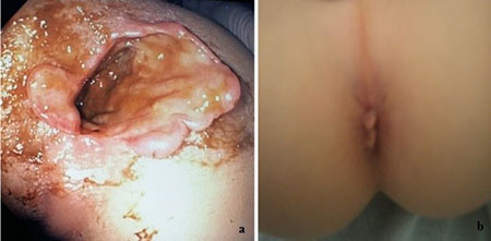
unremarkable. A rectal examination revealed painful inflammation
purulent discharge and stellate lacerations of the anal mucosa and skin
(Fig. 1a).
 |
|
Fig. 1 Perianal abscess of the
3.5-year-old child (a) on admission and (b) 10 days after
treatment.
|
Laboratory investigation upon admission revealed
white blood cell count of 14.9×10⁹/L (neutrophils: 35.4%, lympho-cytes:
55.8%, monocytes: 8.3%) with normal hemoglobin and increased platelet
count. Both C-reactive protein and ESR were mildly raised. Liver and
renal function tests were normal. There was a family history of
recurrent abscesses in mother and maternal aunt raising the suspicion of
immunodeficiency disorder, but all immunological investigation came out
to be normal including classes and subclasses of the immunoglobulins,
immunophenotyping, dihydrorhodamine (DHR) test and cell adhesion
molecules (CAMs). Physical examination findings also raised the
possibility of sexual abuse, which was further ruled out after
behavioral and psychological assessment of child and his parents. The
family was daily reviewed by the pediatric team who found no evidence of
child abuse or family conflicts. There was no evidence of any behavioral
changes or psychological problems in the child during hospitalization
and the follow-up consultation for the next 2 years showed no indication
of psychological problems or any other changes in the behavior of the
child.
Though the stool culture was negative, purulent
discharge culture revealed Klebsiella oxytoca and Proteus
mirabilis and the culture of perianal skin revealed Klebsiella
oxytoca, Enterobacter cloacae and Escherichia coli. The
patient was seronegative for HSV 1 and 2. Rectosigmoidoscopy was
unremarkable and pathology did not reveal any evidence of underlying
inflammatory bowel disease; colonic biopsy revealed only moderate
alterations suggestive of active focal erosive rectitis. Additional
investigations with polymerase chain-reaction in the blood, skin lesion
and rectal tissue for Herpes viruses (HSV1, HSV2, VZV, EBV, CMV)
were also negative. Empiric antimicrobial treatment with cefotaxime,
clindamycin, metronidazole and acyclovir was initiated and continued for
14 days. The clinical course was favorable with complete clinical
resolution (Fig. 1b). In the follow-up period for next two
years, child continued to remain well with no stool incontinence.
We report this unusual case because of the clinical
presentation mimicking lesions associated with sexual abuse, as stellate
lacerations were present. We elected to treat with broad spectrum
antibiotics and provide antiviral treatment. The complete resolution of
his lesions and anal incompetence was remarkable. Since the
investigation did not identify any underlying disease, we concluded that
the most likely pathogenetic cause was the development of severe
neutropenia post viral infection. This highlights the importance of a
complete blood count and a peripheral blood smear in the initial
evaluation of perianal abscess upon presentation. Moreover, although his
family history strongly suggested possible phagocytic dysfunction, the
investigation failed to diagnose such an immune deficiency.
Contributors: DD, AD, SF: conceptualized
the study, drafted the initial manuscript, and reviewed and revised the
manuscript; DD, AD: designed the data collection instruments, collected
data, carried out the initial analyses, and reviewed the manuscript. NZ,
VP: conceptualized and designed the study, coordinated and supervised
data collection, and critically reviewed the manuscript for important
intellectual content. All authors approved the final manuscript as
submitted and agree to be accountable for all aspects of the work.
Funding: None; Competing interest: None
stated.
REFERENCES
1. Fitzgerald RJ, Harding B, Ryan W. Fistula-in-ano
in child-hood: Congenital etiology. J Pediatr Surg. 1985; 20:80-1.
2. Chang HK, Ryu JG, Oh JT. Clinical characteristics
and treatment of perianal abscess and fistula-in-ano in infants. J
Pediatr Surg. 2010;45:1832-6.
3. Ezer SS, Oguzkurt P, Ince E, Hiçsönmez A. Perianal
abscess and fistula-in-ano in children: Etiology, management and
outcome. J Paediatr Child Health. 2010;46:92-5.
4. Whiteford MH. Perianal abscess/fistula disease.
Clin Colon Rectal Surg. 2007;20:102-9.
5. Brook I, Frazier EH. The aerobic and anaerobic bacteriology of
perirectal abscesses. J Clin Microbiol. 1997; 35:2974-6.

