|
|
|
Indian Pediatr 2015;52: 979 -980 |
 |
Mucoepidermoid Carcinoma of Parotid as a
Second Malignancy in Acute Lymphoblastic Leukemia
|
|
*Gargi Tikku, Dhruv Jain, #Archana
Kumari and Rajesh Grover
From the Departments of *Oncopathology, #Radiology
and Radiotherapy, Delhi State Cancer Institute, Delhi, India.
Correspondence to: Dr Gargi Tikku, Department of Oncopathology, Delhi
State Cancer Institute, Delhi, India.
Email: [email protected]
Received: May 02, 2015;
Initial review: May 14, 2015;
Accepted:August 19, 2015.
|
|
Background: Improved survival seen in Acute Lymphoblastic
Leukemia (ALL) cases has led to increased reports of second malignant
neoplasms. Case characteristics: A 12-year-old female treated for
ALL using UK ALL XI protocol nine years back presented with
progressively increasing pre-auricular swelling. Observation:
Investigations revealed it to be a Mucoepidermoid carcinoma. Message:
Mucoepidermoid carcinoma should be a differential in any parotid
swelling of treated case of pediatric ALL.
Keywords: Complication. Recurrence, Second
Malignant Neoplasm.
|
|
T
he most common Second Malignant Neoplasm (SMN)
developing after Acute Lymphoblastic Leukemia (ALL) are central nervous
system (CNS) tumors followed by Acute myeloid leukemia/Myelodysplastic
syndrome [1, 2]. Some reports of SMN in form of salivary
gland Mucoepidermoid carcinoma (MEC) in treated ALL patients have been
published. We report development of the same in an ALL case treated by
United Kingdom ALL XI (UK ALL XI) protocol, which does not entail CNS
radiation as a form of CNS prophylaxis.
Case Report
A 12-year-old female who was a treated case of B
precursor ALL (9 years back), presented with progressively increasing
pre-auricular swelling for 15 days. Examination revealed that the
swelling arose from parotid and was not associated with facial nerve
palsy.
Fine Needle Aspiration Cytology revealed cellular
smears with oval to elongated cells having bland nuclei lying singly and
in groups in a myxoid/mucin rich background. A possibility of a
pleomorphic adenoma was suggested (Fig. 1a).
Contrast-enhanced computed tomography (CECT) of Face and Neck showed an
ill-defined heterogeneously enhancing lesion involving both the lobes of
right parotid gland, likely malignant in etiology. (Fig. 1 b
and 1 c). Additionally, on PET CECT Scan, an FDG -
avid heterogenous solid cystic parotid mass measuring 3.4x2.8 cm was
seen.
 |
|
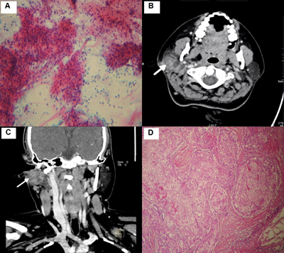
Fig. 1 (a) Cytology smear shows oval
to elongated cells with bland nuclei lying singly and in groups
in a mucin rich/myxoid background. Pap stain (10x) (b) Axial and
(c) coronal CECT image showing heterogeneously enhancing lesion
involving both the lobes of right parotid gland. (d) nests of
mucus and intermediate cells lying in pools of mucin. H & E
(10x).
|
Treatment records showed that standard UK ALL XI
protocol chemotherapy (1992) was administered without any cranial
irradiation. Instead, intravenous and intrathecal Methotrexate along
with folinic acid rescue was given for CNS prophylaxis. Patient remained
in complete remission throughout treatment duration of 3 years and the
bone marrow was in remission during the present admission also.
Right total parotidectomy with facial nerve
end-to-end anastomosis was performed. The pathological diagnosis was
Mucoepidermoid Carcinoma, Grade II. The histology section showed tumor
nests composed of mucus, intermediate and squamoid cells in variable
combinations lying in pools of mucin (Fig. 1d). All
resected lymph nodes were free of tumor. Margins could not be
ascertained as the tumor was removed piecemeal.
Repeat CECT of face and neck, five weeks post-surgery
revealed a residual heterogeneously enhancing tumor in right parotid
region, with invasion of the right temporal bone posteromedially.
Multidisciplinary team decided against surgical intervention and opted
instead for radiation therapy with Intensity Modulated Radiation Therapy
(IMRT) technique. The patient received a total dose of 54 Gy over a
period of one-and-half months. She remains disease-free, 26 months
post-surgery and 23 months post-radiotherapy.
Discussion
Mucoepidermoid carcinoma developing as a second
malignant neoplasm is uncommon with only 19 cases reported following
successful treatment of childhood ALL [1,3-10] (Web Table I).
It was found that the most common site was parotid gland, with only a
single case developing from minor salivary gland of cheek. The MEC
occurred as the second malignancy, 8.1 years (mean) after the initial
leukemia diagnosis.
All previously reported 19 patients diagnosed as ALL
were treated by multi-drug chemotherapy along with either intrathecal
methotrexate and prednisolone or cranial irradiation. Eight out of these
19 cases, had received MDC along with cranial irradiation, and most had
received a dose of 18Gy [1,3,6,9,10]. Total Body Irradiation was also
given in another three cases along with MDC [5, 9]. Six cases had
received multi-drug chemotherapy without any radiotherapy [4,7,9,]. This
is in keeping with our case, highlighting possible role of MDC, in
addition to the known role of radiation in development of MEC in treated
ALL cases. The risk factors for development of second malignant
neoplasm. include radiation to the craniospinal axis, ALL with CNS
involvement, relapse of primary disease, female sex and
epipodophyllotoxins as frontline agent [1,2]. Our case exhibited the
latter two risk factors, with etoposide having been administered.
Cyclophosphamide use has also been proposed as a risk factor [9], but
without adequate evidence.
Mucoepidermoid carcinoma are usually low grade, and
misdiagnosed on FNAC
[3,4,6-9]. Most reported patients were
alive and free of disease after treatment of MEC regardless of
histological grade [1,3-9]. This reinforces the importance of timely and
correct diagnosis of this malignant neoplasm so that early surgical
treatment can be instituted.
To conclude, mucoepidermoid carcinoma even though
rare, should always be kept as a differential in any parotid region
swelling of a treated case of pediatric ALL as these SMN’s are mostly
low grade and very much amenable to treatment thereby increasing the
chances of survival in young patients.
Contributors: All authors participated in the
management of the patient and drafting of manuscript. The final version
was approved by all authors.
Funding: None; Competing interests:
None stated.
References
1. Bhatia S, Sather HN, Pabustan OB, Trigg ME, Gaynon
PS, Robison LL. Low incidence of second neoplasms among children
diagnosed with acute lymphoblastic leukemia after 1983. Blood.
2002;99:4257-64.
2. Loning L, Zimmermann M, Reiter A, Kaatsch P, Henze
G, Riehm H, et al. Secondary neoplasms subsequent to
Berlin–Frankfurt–Munster therapy of acute lymphoblastic leukemia in
childhood: Significantly lower risk without cranial radiotherapy. Blood.
2000;95:2770-75.
3. Loy TS, McLaughlin R, Odom LF, Dehner LP.
Mucoepidermoid carcinoma of the parotid as a second malignant neoplasm
in children. Cancer. 1989;64:2174-77.
4. Kaste SC, Hedlund G, Pratt CB. Malignant parotid
tumors in patients previously treated for childhood cancer: clinical and
imaging findings in eight cases. AJR Am J Roentgenol. 1994;162:655-59.
5. Atahan IL, Ayhan A, Ozyar E, Ertoy D, Gürkaynak M.
A case of mucoepidermoid carcinoma of the parotid gland developing in a
child after the treatment of acute lymphoblastic leukemia. Pediatr
Hematol Oncol. 1995;12:403-05.
6. Myer CM III. Second primary malignancies of the
head and neck in children. Am J Otolaryngol. 1995;16:415-7.
7. Sandoval C, Jayabose S. Parotid
mucoepidermoid carcinoma following chemotherapy for childhood acute
lymphoblastic leukemia. Pediatr Hematol Oncol. 2001;18:217-20.
8. Savelli SL, Klopfenstein KJ, Termuhlen AM.
Mucoepidermoid carcinoma of the parotid gland as a second malignant
neoplasm. Pediatr Blood Cancer. 2005;45:997-1000.
9. Védrine PO, Coffinet L, Temam S, Montagne
K, Lapeyre M, Oberlin O, et al. Mucoepidermoid carcinoma of
salivary glands in the pediatric age group: 18 clinical cases, including
11 second malignant neoplasms. Head Neck. 2006;28:827-33.
10. Tugcu D, Akici F, Aydogan G, Salcioglu Z,
Akcay A, Sen H, et al. Mucoepidermoid carcinoma of the parotid
gland in childhood survivor of acute lymphoblastic leukemia with need of
radiotherapy for treatment and review of the literature. Pediatr Hematol
Oncol. 2012;29:380-85.
|
|
|
 |
|
