|
|
|
Indian Pediatr 2014;51:
921-923 |
 |
Radial Artery Pseudoaneurysm in a Neonate with
Hemophilia A
|
|
Shrenik Vora, Thowfique Ibrahim And Victor Samuel
Rajadurai
From Department of Neonatology, KK Women's and
Children's Hospital, 100, Bukit Timah Road, Singapore.
Correspondence to: Dr Shrenik Vora, Staff Registrar,
Department of Neonatology, KK Women's and Children's Hospital, 100,
Bukit Timah Road, Singapore 229899,
Email:
[email protected]
Received: March 24, 2014;
Initial review: June 30, 2014;
Accepted: September 02, 2014.
|
|
Background: Pseudoaneurysm
formation is a rare complication of arterial puncture. Case
characteristics: 3-week-old male developed an enlarging mass over
the anterior aspect of left wrist following radial arterial puncture.
Observation: Doppler ultrasonography revealed mass to be left radial
arterial pseudoaneurysm. Subsequent presentation of ecchymoses and
investigations confirmed factor VIII deficiency (Hemophilia A).
Outcome: Pseudoaneurysm removed with primary end to end anastomosis.
Patient is presently on regular factor VIII replacement therapy.
Message: Hemophilia A can present as pseudoaneurysm in neonatal
period.
Keywords: Arterial cannulation, Complications,
Factor VIII.
|
|
Trauma to the wall of an artery can lead to the
formation of pseudoaneurysm, a painful expanding mass overlying the
damaged vessel[1]. 30% of severe factor VIII deficiency presents in
neonatal period [2]. There are infrequent case reports of hemophilia
presenting as pseudoaneurysm in children, more so in neonates [3]. We
describe a case report of radial artery pseudoaneurysm following
arterial puncture in a newborn infant with hemophilia A.
Case Report
A 3-week-old large-for-gestational age male neonate
admitted for presumed sepsis, presented with an enlarging mass at the
anterior aspect of left wrist. A week prior to this, an arterial
puncture with 24-guage needle had been attempted at the site for blood
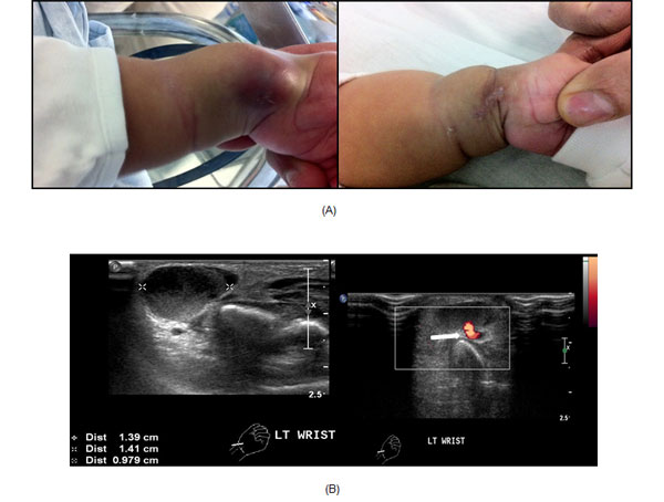
collection. Physical examination showed a 1×1 cm, firm, non-pulsatile,
non-warm swelling over left radial artery, with good capillary refill of
distal upper extremity (Fig.1a). Doppler ultrasonography
revealed 1.4×1.4×1 cm cystic lesion with moving internal echoes situated
adjacent to left radial artery with small connection with the artery
suggestive of left radial arterial pseudoaneurysm (Fig. 1b).
The mass progressively increased in size and the patient underwent an
operative repair consisting of excision of pseudoaneurysm with primary
end to end anastomosis. There were three puncture marks over left radial
artery with no evidence of inflammation or infection and there was no
difficulty in achieving intra-operative hemostasis. Post-operatively,
palpable pulses and good doppler flow were established in the left
radial and ulnar artery.
 |
|
Fig.1 (a) Left radial artery
pseudoaneurysm- pre- and post- repair; and (b) Sonogram showing
cystic lesion with doppler color flow image showing internal
echoes (white arrow).
|
The child subsequently developed spontaneous bruises
and ecchymoses at lower limbs. The activated partial thromboplastin time
was prolonged (>180 secs) and diagnosis was confirmed as hemophilia A
(factor VIII levels < 1%). Factor IX, XI, XII, von-willebrand factor and
ristocetin factor levels were in acceptable range. Mother's activated
partial thromboplastin time and factor VIII levels were within normal
range, and there was no family history of hemophilia. Post-discharge,
the child was started on regular factor VIII replacement therapy. On
follow-up at one year of age, clinical examination revealed palpable
left radial pulses and normal growth of the left upper extremity without
any neurological deficit.
Discussion
Arterial trauma may lead to occlusion of the vessel
by thrombosis, development of arterio-venous fistula or the formation of
an arterial pseudoaneurysm [4]. Pseudoaneurysm formation is a delayed
complication arising due to disruption of the vessel wall with
containment of blood by surrounding tissues and appearance of sac in
direct continuity with arterial lumen. The most common cause of
pseudoaneurysm is blunt or penetrating trauma to artery from cannulation
or puncture, while less common etiologies are connective tissue
disorders, bleeding tendencies, infection and inflammation [5]. Severe
factor VIII deficiency mainly presents in neonatal period as excessive
hematomas, post-delivery cephalhematomas, post-surgical bleeding and
intracranial bleed [2]. Pseudoaneurysm in hemophilia is known, but there
has been a limited description of bleeding disorders presenting as
arterial pseudoaneurysm in the neonatal period [6].
Most common means of presentation of pseudoaneurysm
is that of a palpable pulsatile or non-pulsatile expanding mass with
palpable distal pulses and a vital extremity. Pressure applied to the
mass will result in decompression of the blood filled sac whereas
release leads to rapid refill, thereby distinguishing from a solid mass
and it should be carefully examined for a palpable thrill and audible
bruit. An effort should always be made to look at other diagnosis like
connective tissue disorder, coagulopathy or arteritis [7]. In our case,
the appearance of spontaneous bruises and ecchymosis along with the
formation of left radial artery pseudoaneurysm lead us to investigate
further and confirm the diagnosis of bleeding diathesis. Doppler
sonography or CT scan is required to assess the presence of vascular
flow within the mass and to differentiate the lesion from either a tumor
or abscess [8]. Traditionally arterial pseudoaneurysms are managed in
various ways: observation, compression bandages, ultrasound-guided
compression, ultrasound-guided thrombin injection, and surgical repair
[9]. Factors requiring urgent surgery include bleeding, evidence of
vascular compromise or increasing size of the pseudoaneurysm (as in our
case). Patients with arterial pseudoaneurysm need to be followed up for
a prolonged period to assess possible development of growth
disturbances, neurological deficit, and limb length discrepancies [10].
To conclude, radial artery cannulation being a very
common procedure in clinical practice for arterial blood gas analysis
and cardiovascular monitoring in neonates and infants, physicians and
nurses in pediatrics units should be aware of the possibility of
incurring arterial injuries, necessitating early diagnosis and prompt
intervention of pseudoaneurysms. Though a small-caliber needle was used
for radial arterial puncture in our case, his underlying bleeding
diathesis contributed to the formation of radial arterial pseudoaneurysm.
One should be vigilant to rule out other rarer causes of pseudoaneurysm
like bleeding disorders, inflammation and infection.
Acknowledgement: Dr Por YC, for his help in the
surgical management of the patient.
Contributors: SV: Literature review, manuscript
drafting, review and editing, and patient management; TI: Literature
review, manuscript editing and patient management; VSR: Guidance and
final editing of manuscript with extensive literature search. All
authors approved the final manuscript.
Funding: None; Competing interests: None
stated.
References
1. Ganchi PA, Wilhelmi BJ, Fujita K, Lee WP. Ruptured
pseudoaneurysm complicating an infected radial artery catheter: case
report and review of the literature. Ann Plast Surg. 2001;46:647-50.
2. Chalmers E, Williams M, Brennand J, Liesner R,
Collins P, Richards M. Guideline on the management of hemophilia in the
fetus and neonate. Br J Haematol. 2011;154:208-15.
3. Rodriguez-Merchan EC. Pseudoaneurysms in
haemophilia. Blood Coagul Fibrinolysis. 2013;24:461-4.
4. Rey C, Manache P, Watel A. Iatrogenic false
aneurysm of the brachial artery in an infant. Eur J Pediatr.
1987;146:438-9.
5. Ivo D, Josip U, Davor M, Kresimir B.
Pseudoaneurysms of the brachial artery following venipuncture in
infants. Pediatr Surg Int. 2004;20:594-7.
6. Fields JM, Saluja S, Schwartz DS, Touloukian RJ,
Keller MS. Hemophilia presenting in an infant as a radial artery
pseudoaneurysm following arterial puncture. Pediatr Radiol.
1997;27:763-4.
7. Kenneth WG, James M, Ellen LP, Thomas FD. Brachial
artery pseudoaneurysm in a 6-week-old infant. Amer Surg. 2004;06:518-21.
8. Restrepo R, Ranson M, Chait PG. Extracranial
aneurysms in children: Practical classification and correlative imaging.
AJR. 2003;181:867-78.
9. Landau D, Schreiber R, Szendro G, Golcman L.
Brachial artery pseudoaneurysm in a premature infant. Arch Dis Child
Fetal Neonatal Ed. 2003;88:F152-3.
10. Wolffgang BG, Steven MS, Todd DR. Radial artery
pseudoaneurysm in the intensive care unit. Ann Vasc Surg.
2010;24:554e13-16.
|
|
|
 |
|
