|
|
|
Indian Pediatr 2013;50: 1073-1074 |
 |
Childhood Bullous Mastocytosis
|
|
Dipti Das, Anupam Das and Swapan Sardar
Department of Dermatology, Medical College and
Hospital, Kolkata, West Bengal, India.
Email: [email protected] m
|
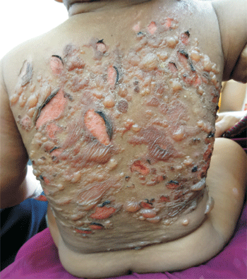
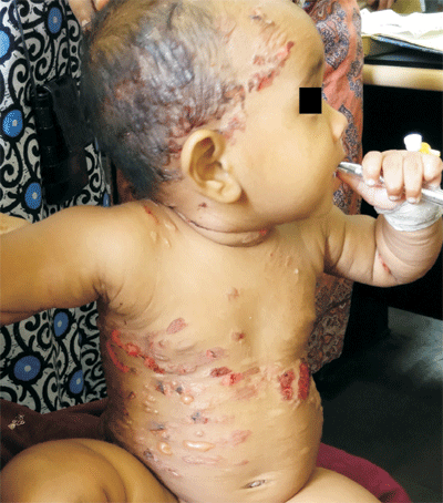
A 1-year-old girl presented with numerous pruritic bullae
all over the body. There were complaints of recurrent
episodes of diarrhea and vomiting. Development was normal.
She started developing episodes of intense itching at 8
months of age, followed by appearance of erythematous
macules, plaques, and tense bullae at the sites of itching
as well as other sites (scalp, trunk and upper extremities)
(Fig. 1 and 2). Bullae were present on a non-urticated
base containing clear fluid. Few of the urticarial plaques
showed peau-d-orange appearance. Darier’s sign was
positive. The palms, soles and mucosae were free. Routine
investigations and urine analysis were normal. Skin biopsy
showed sub-epidermal bulla and an upper dermal inflammatory
infiltrate comprising lymphocytes and many mast cells.
Toluidine blue staining showed metachromatic granules and a
diagnosis of bullous mastocytosis was made. The patient was
treated with antihistamines for itching and topical as well
as systemic antibiotics for preventing secondary infection.
Parents were counseled regarding the prognosis and course of
disease and the importance of avoiding certain medications
that may provoke mast cell degranulation.
 |
 |
|
Fig. 1 Multiple tense
bullae and erosions over the trunk.
|
Fig.2 Bullae, plaques and
erosions on face and trunk.
|
Bullous mastocytosis is a severe variant
of mastocytosis, 30% cases manifesting within 6 months of
age. The typical childhood disease is linked to Glu-839-Lyc
c-kit mutation. Commonest clinical variants are
telangiectasia macularis eruptiva perstans, mastocytoma,
diffuse cutaneous mastocytosis and urticaria pigmentosa.
Many agents stimulate the degranulation of mast cells, such
as bacterial toxins, physical stimuli, poisons, biological
peptides, polymers and drugs like aspirin, codeine,
morphine, quinine etc. The close clinical differentials
include chronic bullous disease of childhood, epidermolysis
bullosa and staphylococcal scalded skin syndrome (SSSS).
Combinations of H1 and H2 blocking agents have been the
mainstay of treatment for most of the uncomplicated cases.
|
|
|
 |
|
