An 11-year-old boy presented with raised pigmented lesion
over the scalp since 10 years. Parents gave history of
hairless yellowish plaque present over the scalp at birth
which gradually increased to present size to take
cerebriform appearance. There was no history of trauma. All
routine hematological investigations were normal. No
systemic and developmental defect was noted. X-ray
skull, eye and neurological examination were normal.
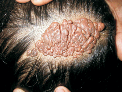
On cutaneous examination, single, 17×8 cm
brownish, soft, cerebriform and well demarcated nodular
plaques was present over the scalp (Fig. 1).
It had multiple folds. Histopathology showed marked
papillomatous epidermal hyperplesia with hyperkeratosis and
large numbers of mature sebaceous glands in the dermis along
with follicular plugging with malformed hair follicles were
also present. The correlation diagnosis of cerebriform type
of nevus sebaceous was made.
 |
|
Fig.1 Cerebriform nevus
sebaceous.
|
Nevus sebaceous of Jadassohn (NS) is an
epidermal nevus, predominantly congenital sebaceous
hamartoma with an estimated incidence of 0.3% in the
neonates. PTCH gene deletion is proposed mechanism for
development of nevus sebaceous. Cerebriform type is a very
rare morphologic variant of NS.
It is usually located over head and neck
region as solitary lesion and often present at birth as
single hairless yellowish plaque with a smooth velvety
surface. Multiple extensive lesions may develop with linear,
blaschkoid pattern. It becomes verrucous and nodular at
puberty indicating role of hormones. Common sites are scalp,
forehead, centrofacial, periauricular, and genital area. It
may be associated with other developmental defects which are
included as epidermal nevus syndrome. Though it occurs
sporadically, autosomal dominant transmission was suggested
by many case reports. Trichoblastoma is most common benign
tumor which develops secondarily in NS, while malignant
tumor is basal cell carcinoma (<5%).
The clinical differential diagnosis is
congenital melanocytic nevi, epidermal nevus syndrome, giant
seborrheic keratosis and warts, while the histopathological
differential diagnosis is sebaceous hyperplesia, adenoma,
sebaceous carcinoma and sebaceoma.
Seborrheic keratosis and epidermal
nevus may be difficult to differentiate clinically.
Sebaceous adenomas is sharply demarcated structure made up
of immature lobules while in sebaceoma basaloid cells
predominate along with sebaceous cells ducts. In contrast to
nevus sebaceous, sebaceous hyperplasia shows mature
sebaceous gland lobules and prominent sebaceous ductal
structures. Sebaceous carcinoma shows mitotic cells with
undifferentiated growth.
Wide excision remains treatment of choice
and patient mainly present for cosmetic purpose. It may be
done prophylactically during childhood as there is a risk of
malignant transformation, but most tumors remain benign. So
regular clinical follow up is necessary. Many other
treatment modalities like CO2 laser and photodynamic therapy
have been tried.


