|
|
|
Indian Pediatr 2012;49: 936-937 |
 |
Pityriasis Lichenoides
|
|
Shylaja Someshwar and Satish Udare
Department of Dermatology, MGM Medical College, Navi
Mumbai, Maharashtra, India.
Email: [email protected] n
|
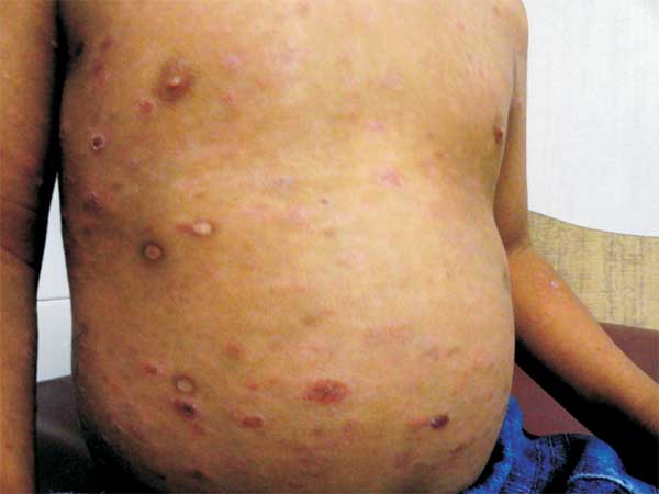
A 2-year-old male child presented with multiple asymptomatic
erythematous scaly papules and plaques with peripheral
scales all over the face, scalp, trunk and extremities since
3 weeks (Fig.1). Prior to this, there was a
history of fluid filled lesions of 2 weeks duration which
left behind Chicken pox like scars following symptomatic
treatment. Skin biopsy showed typical features of Pityriasis
lichenoides et varioliformis acuta (PLEVA). He was treated
with narrow band UVB with good results.
 |
|
Fig. 1 Erythematous scaly
lesions and scars.
|
Clinical spectrum of Pityriasis
lichenoides ranges from acute papular lesions that rapidly
evolve into papulovesicles, necrosis and varioliform
scarring (PLEVA) to small, scaly, benign-appearing papules (pityriasis
lichenoides chronica or PLC) both with a generalized
distribution. This affects young adults and occasionally
children. Though the etiology of this condition is unclear,
infectious/drug related hypersensitivity and premycotic
lymphoproliferative disorder are the mainstay theories. The
diagnosis is by histopathology. In
PLEVA, as there are polymorphic lesions of papules and
vesicles appearing in crops which may heal with scars, a
differential diagnosis of varicella has to be considered.
Hemorrhagic necrosis and a course of waxing and waning are
not common in varicella.
As PLC presents as small papules with
scaling, pityriasis rosea and psoriasis need to be ruled
out. While typical pattern and a self-limiting benign course
excludes pityriasis rosea; symmetry, distribution pattern
and silvery scales point to the diagnosis of psoriasis. PLC
may also have adherent ‘mica like’ scales, which, when
detached, reveal a shiny brown surface. If seen, this is a
distinctive diagnostic feature. The present case had
features of both PLEVA and PLC.
Though the condition is self-limiting, as
the course is unpredictable, it warrants therapy. Oral
antibiotics, topical corticosteroids and phototherapy have
been tried with variable success.
|
|
|
 |
|
