|
|
|
Indian Pediatr 2012;49: 935 |
 |
Hyperkeratotic Scaly Lesions
|
|
Piyush Kumar, Vikas Anand and Sambeet Kumar Mallik
Department of Dermatology, Katihar Medical College,
Katihar, Bihar, India.
Email: docpiyush@gmail.com
|
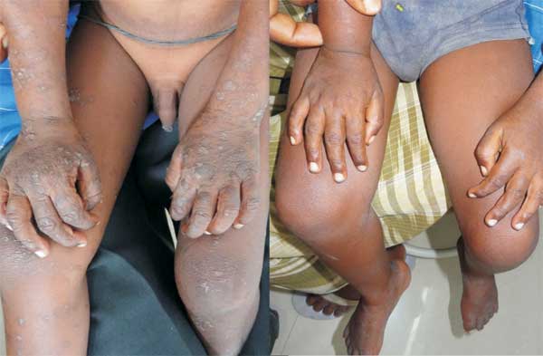
A 4-year-old boy presented with mildly itchy hyperkeratotic
scaly lesions on pressure points like knees, elbows,
buttocks and ankles in a bilateral symmetrical manner for
two months (Fig. 1). Parents also complained
of recent onset lethargy and diarrhea for three weeks. Child
and parents both were known to be HIV- seropositive and were
on anti-retroviral therapy. Rest of the mucocutaneous
examination was unremarkable, except for oral candidiasis.
Tinea incognito, atopic dermatitis and acquired zinc
deficiency were considered as differential diagnoses. In
atopic dermatitis, the lesions are itchy, predominantly over
flexors (on extensors in infants) and are accompanied by
other features e.g. hyperlinearity of palms, Dannie
Morgan folds, generalized xerosis, follicular papules, and
history of atopy. These findings were absent in our patient.
HIV status of baby, asymptomatic psoriasiform scaly plaques
over pressure/trauma prone areas, absence of features of
inflammation and concurrent diarrhea were clinical clues for
a diagnosis of acquired hypozincemia in a HIV patient. KOH
mount from the scaly lesions were negative for fungus and
histopathology revealed psoriasiform acanthosis, confluent
parakeratosis and mild neutrophilic infiltration. The
epidermis was notable for pallor of upper layers and few
dyskeratotic keratinocytes. These findings were consistent
with the diagnosis of acquired hypozincemia. Serum zinc
level could not be estimated, but alkaline phosphatase level
was marginally low (82 IU/L). He was treated with oral zinc
at a dose of 2 mg/kg/day and emollient (and clotrimazole
mouth paint for oral candidiasis). On follow up visit after
two weeks, the cutaneous lesions and diarrhea were
completely improved (Fig. 1). The parents were
counseled about including zinc-rich food items in diet.
 |
|
(a)
(b) |
|
Fig. 1 (a) Dry
hyperkeratotic scaly lesions on pressure points
(left). Note absence of crural region
(characteristically involved in Acrodermatitis
enteropathica), (b) complete clearance of lesions
within two weeks.
|
The clinical manifestations of acquired
zinc deficiency are variable and depend on degree of
hypozincemia. The patients with significant hypozincemia
present with periorificial erosive dermatitis, mimicking
acrodermatitis enteropathica (AE). However, patients with
mild hypozincemia present with less dramatic, less
characteristic psoriasiform lesions which may be missed by
treating physicians. In mild hypozincemia, lesions are
characterized by "scald like" erythema and lack of
inflammation. The lesions are typically seen on areas
subject to repeated pressure and trauma, such as elbows,
knees, knuckles, ankles, and sacral area. The cutaneous
lesions may be psoriasiform plaques (in appearance and
distribution), annular plaques with brown black crusts at
advancing margins, crusted plaques, or pigmented lichenoid
lesions. Vesicobullae, pustules, and erosions are occasional
findings. There is decreased hair and nail growth. The
diagnosis of mild hypozincemia requires a high index of
suspicion. The determination of a low plasma zinc level is
diagnostic (normal 70-250 µg/dL), but may not be helpful in
all cases as it suffers from many limitations. Serum
alkaline phosphatase and histopathology may aid in
diagnosis. The clinical response to zinc supplementation
(1-2 mg/kg/day) remains the gold standard of diagnosis.
Typically, there is rapid improvement of diarrhea within 24
hours and skin lesions heal within 1 to 2 weeks of zinc
therapy without additional topical therapy.
|
|
|
 |
|
