|
|
Case Reports Indian Pediatrics 2007;44:861-863 |
||
|
Bullous Systemic Lupus Erythematosus and Lupus Nephritis in a 10-year-old Boy |
||
|
A.M. Vijayalakshmi From the Department of Pediatrics, PSG. Institute of Medical Sciences and Research, Coimbatore 641 004, India. Correspondence to: Dr. A.M. Vijayalakshmi, Department of Pediatrics, PSGIMS & R, Coimbatore, Tamil Nadu, India. E-mail: [email protected]
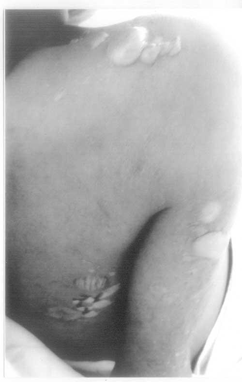
Abstract: Key words: Bullous systemic lupus erythematosus, Class V lupus nephritis, Nephrotic syndrome. Bullous systemic lupus erythematosus (BSLE) is an autoantibody mediated subepidermal blistering disease that occurs in patients with SLE. It is characterized by distinctive combination of clinical, histologic and immunopathologic features. Bullous SLE manifests in the second through third decades of life. It is very rare in children with only five cases published, most of them girls(1). We report a 10-year-old boy with bullous SLE and lupus nephritis. This is the first case reported at such young age in a literature search. Case Report A 10-year-old boy presented with intermittent fever for four months, recurrent episodes of bullous lesions of skin for one month and generalised edema for one week. On examination he was febrile, sick looking and had puffiness of face, edema of legs and scrotum. He had ulcers over the hard palate and left cheek. There was no generalized lymphadenopathy, malar rash or discoid rash. He had tense bullae of varying sizes over the ears, lateral aspect of the neck, shoulders, arms and legs (Fig. 1). The skin lesions started as vesicles which progressed to become bullae within 2 days. The blisters ruptured spontaneously to form erosions and then crusting followed by depigmentation. His hair was sparse and nails were normal. Examination of cardio-vascular system revealed no evidence of heart failure, cardiomegaly or conduction abnormalities. Examination of respiratory system showed features of bilateral pleural effusion. His abdomen was distended with free fluid and liver was palpable 3 cm below right costal margin and spleen was not palpable. He had hypertension with the BP of 130/ 90 mm of Hg. His ophthalmic examination including fundus was normal.
Investigation revealed total WBC count of 4,900/cu mm, Hb of 9.5 g/dL, RBC count of 3.15 millions / µL platelet count of 4,85,000/cu mm, ESR of 105 mm/h and normocytic, normochromic anemia. Urine analysis showed 4+albumin, 45-50 RBC / hpf, 55-60 WBC / hpf, 3-5 granular casts and 4-5 hyaline casts / hpf. Mantoux test was negative and X-ray chest showed bilateral pleural effusion. Blood urea was 21 mg / dL and serum creatinine 0.6 mg/dL. Total serum protein was 5.6 g/dL, albumin 1.5 g/dL and globulin 4.1 g/dL, serum cholesterol was 313 mg/dL. Serum elctrolytes showed sodium 138 mEq/L, potassium 3.76 mEq/L, chloride 102 mEq/L, bicarbonate 20 mEq/L. Blood culture was sterile and urine culture had grown 10,000 colonies / mL of E.coli. Antinuclear antibody by ELISA was 4 (>1 positive) and antibodies to double stranded DNA was 3.7 (>1 positive) by EUROIMMUN kit. His lupus anti-coagulant and anti-cardiolipin antibodies were negative. Ultra-sound showed moderate ascitis, mildly enlarged kidneys and bilateral pleural effusion. Histopathological study of skin biopsy revealed subepidermal blister with neutrophilic dermal infiltration. Direct immunoflorescence study of skin biopsy specimen showed strong granular basement membrane zone band of IgG and moderately strong granular basement membrane band of IgM, IgA and C3 suggestive of bullous SLE. Renal biopsy specimen showed features of membranous nephropathy suggestive of Class V lupus nephritis. Since the child presented with prolonged fever, nephrotic syndrome and skin lesions, HIV was considered in the differential diagnosis but HIV routine ELISA was non-reactive for HIV 1 and HIV2 antibodies.Based on the clinical features and investigations a diagnosis of bullous SLE with lupus nephritis was made. He was started on oral prednisolone 2 mg/kg/day dapsone 100 mg/day and losartan 50 mg/day. Within 4 weeks of above treatment, his edema resolved, bullous lesions healed completely leaving depigmented areas and he became normotensive. After 2 months of daily steroid therapy was over, an attempt was made to taper steroid. When prednisolone was tapered to 0.5 mg/kg/day he developed erythematous patches and painful swelling of both soles suggestive of vasculitis and was unable to walk. Prednisolone was increased to 2 mg/kg again and the vasculitis resolved with in a week. He has been on close follow up for the last 12 months. Prednisolone, dapsone and losartan were stopped after 10 months from onset. At recovery the repeat ESR was 11 mm/hr, ANA level was 7.979 (positive) and DS DNA level was 0.240 (negative). Without any medication the child is doing well for the last 2 months and there is no recurrence of bullous skin lesions or relapse of nephrotic syndrome. Discussion Bullous SLE is an extremely uncommon subset of systemic lupus erythematosus with an incidence of 0.26 per million population per year in adults(2). Clinically, BSLE is characterized by rapid develop-ment of widespread vesiculobullous lesions. Blisters range from large tense bullae to small vesicles. Bullae contain clear or hemorrhagic fluid and rupture spontaneously resulting in erosion and crusts which heal with hypopigmentation or hyperpigmentation. The blisters are distributed over the neck, face, trunk and extremities. Among the cutaneous manifes-tations of SLE, BSLE accounted for 2 percent in THAI children with SLE(3). Rarely, BSLE may be the initial clinical manifestation of SLE(4,5). Though, bullous skin lesions were the initial manifestation of SLE in our case, he had features of nephrotic syndrome also at admission. Bullous SLE presenting in a localized linear pattern has been reported(6). The histological examination in BSLE shows neutrophilic dermal infiltrate and subepidermal separation(7). Direct immunofluore-scence (DIF) of perilesional skin reveals deposition of IgG with or without Ig M and Ig A and complement in a linear or granular bandlike pattern at the basement membrane. Our patient’s skin biopsy has shown typical histologic and DIF features. Class V membranous nephritis occurs in 10% to 20% of patients with lupus nephritis(8). The clinical presentation ranges from mild proteinuria with hematuria to nephrotic range proteinuria. Hypertension occurs in 30% of cases. Our child had Class V lupus nephritis and presented with nephrotic syndrome. The association of lupus nephritis with BSLE has been reported in adults(5). Bullous SLE shows dramatic response to dapsone(5,9). Fifty to 100 mg/day is often used (10). In our child cessation of new blister formation occurred within 48 hours after start of dapsone therapy and existing lesions healed within a week. Most patients with Class V lupus nephritis require only low doses of steroids for a short period(8). Only a minority of patients require a prolonged course of steroids or use of a second immunosuppressive agent like cyclosporin or azathioprine. Our child responded well to oral steroids. Our case illustrates that generalized vesiculobullous eruption can be the initial manifestation of SLE and the close relationship between BSLE and lupus nephritis. Contributors: AMV: diagnosis and management of patient, preparation of manuscript, review of the literature, review of the manuscript; AJ: involved in management of patient and review of literature. Funding: None. Competing interests: None stated. | ||
|
References | ||
|
![]()