|
|
Case Reports Indian Pediatrics 2006;43:991-994 |
||||
|
Fungal Brain Abscesses in Leukemia |
||||
|
Fani Athanassiadou 2nd Pediatric Department, Aristotle University of Thessaloniki, AHEPA Hospital, Thessaloniki, Greece and *Microbiology Laboratory, Athens Medical School, Athens, Greece.
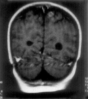
Invasive fungal infections are common in immunocompromised children receiving cytotoxic chemotherapy for hematological malignancies. Invasive central nervous system (CNS) aspergillosis is rare and accounts for about 10% of all cases of invasive aspergillosis(1,2). Cryptococcal CNS infection is uncommon and mainly associated with manifestations of meningitis and encephalitis(3-5). Significant morbidity and mortality are associated with Aspergillus and Cryptococcus CNS infections, and only occasional case reports note survival. Case Report Case 1 A two-year-old boy was referred to our department for fever and pancytopenia. Clinical and laboratory examination revealed evidence of B-ALL and the boy received induction chemotherapy with ALL BFM 95 protocol. One month after initiation of cytotoxic chemotherapy he presented with a febrile neutropenia episode and developed symptoms of upper airway obstruction accompanied by cough. Bronchoalveolar lavage (BAL) culture was positive for Aspergillus fumigatus. PCR confirmed detection of Aspergillus DNA in blood and sputum. Treatment was started with liposomal amphotericin (LAmB) (3 mg/kg/day i.v.). Two weeks after initiation of antifungal treatment the patient had sudden onset of CNS symptoms with headache. Magnetic resonance imaging (MRI) of the brain with contrast revealed the presence of a single lesion in the right parietal lobe showing ring enhancement indicative of abscess (Fig.1). He was immediately treated with high doses of LAmB (10 mg/kg/day i.v.) and voriconazole (VRC) (8 mg/kg/day i.v.). MRI scan performed 6 weeks later showed a nearly total regression of the abscess. At 8 months evaluation, the patient was asymptomatic, CT and MRI scans of chest and brain were normal and antifungal treatment was discontinued. The patient has completed consolidation chemotherapy treatment and is in a good clinical condition.
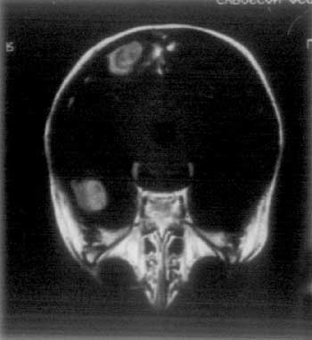
Case 2 A 5-year-old boy was admitted to our department with fever, anemia and cervical lymphadenopathy. Clinical and laboratory examination revealed evidence of B-ALL. The boy was treated with ALL BFM 95 protocol. Six months later, while on reinduction chemotherapy phase, the patient had a sudden onset of CNS symptoms with headache, nausea and vomiting. On clinical examination the boy was afebrile, didn’t have focal neurological deficits or signs of meningeal irritation. Tests for Candida and Aspergillosis were negative, while CSF showed no cells. He was started on empirical antibiotic therapy (ceftriaxone) but two days later, patient’s clinical status worsened. On the basis of a second lumbar puncture result [300/mm3 leukocytes (280 lymphocytes), mild increase in CSF protein levels (181 mg/dL) and hypoglycorrachia (15 mg/dL)] and the ineffectiveness of the antibiotic therapy, he was started on LAmB (5 mg/kg/day) and ceftazidime. Diagnosis of Cryptococcus neoformans infection was based on laboratory exams (serum and CSF antigenic titers by latex agglutination test were 1:101 and 1:10000 respectively, positive detection of cryptococcal DNA by PCR, isolation of the microorganism in the urine). MRI scan findings revealed the presence of two focal lesions indicative of cryptococcoma (Fig. 2). Radiological findings on MRI scan 2 weeks after initiation of LAmB have demonstrated the eradication of the parietal lesion and the permanence of the occipital one. The patient’s clinical status deteriorated rapidly and was admitted in an intensive care unit but unfortunately he died of pulmonary and renal insufficiency 4 weeks later.
Discussion Cerebral aspergillosis occurs in about 10 to 20% of all cases of invasive aspergillosis, and has a very poor prognosis. Case fatality rate (CFR) exceeds 88%, with a value that may be near 100% in many studies(1). The poor prognosis of brain abscesses in immunocompromised patients is well documented in the Fred Hutchinson Cancer Research Center series in which 56 of 58 transplant patients who developed a brain abscess died shortly after diagnosis(2). Signs and symptoms frequently reported in literature include headache, focal neurological deficits and change in mental status. CNS cryptococcosis is mainly associated with manifestations of meningitis and encephalitis such as headache, nausea, confusion, irritability and vision disturbances(3-5). Both fever and nuchal rigidity are usually absent as in our patient. Lumbar puncture (CSF findings similar to that found in tubercular meningitis) and serological antigenic titers are useful diagnostic criteria(6). Many studies in literature have described a series of findings related to poor prognosis such as cortico-steroid treatment, low glucose levels in CSF and high titers of cryptococcal antigen (>1:32), findings compatible with ours(7-8). The spectrum of MRI abnormalities described in CNS cryptococcosis ranges from no abnormality to meningeal enhancement with/or focal single/multiple cryptococcal lesions with defined fibrous capsulae(8). To our knowledge this is the first case of crypto-coccoma caused by Cryptococcus neoformans in a child with acute lymphoblastic leukemia reported in literature. In patients with hematological malignancies brain abscesses on MRI scans have been reported to depend on immunological status of patient(9). Therefore, in patients with severe immunosuppression the brain fungal lesions are consistent with acute infarct and usually are associated with a fatal outcome. This is probably due to the rapid evolution of the disease and absence of inflammatory response. In patients without severe immunosuppression, fungal brain lesions show ring or nodular enhancement consistent with abscess or granuloma. The prognosis of these patients is favorable because of the host’s capacity to isolate or encapsulate the fungal micro-organism. The MRI scans of our children were indicative of patients without severe immuno-suppression. In conclusion, fungal brain abscesses are rare but disabling complications following intensive chemotherapy for childhood ALL. Prognosis of these patients is poor and depends on the early recognition of the causative organism and prompt initiation of antifungal treatment. Contributors: FA managed both cases and prepared the manuscript. AT reviewed the literature and wrote the manuscript. TP managed both cases. AV did laboratory diagnosis of fungal infection. Funding: None. Competing interest: None..
| ||||
|
References | ||||
|
|
![]()