|
|
Case Reports Indian Pediatrics 2002; 39:1047-1050 |
||
|
Urinoma |
||
|
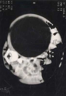
Renal trauma in children is relatively common following blunt abdominal trauma and is usually managed conservatively with good results(1). Rarely extravasation of urine occurs in the perirenal space to form an urinoma. Urinoma may also be found in extraperirenal, retroperitoneal locations, peritoneal cavity, pleural space or the media- stinum. It becomes encapsulated by a very thick fibrous lining to form a mass(2). This is usually a consequence of some fistulous drainage in the urinary tract following injury, surgery or a leak in collecting system(3). We report a case of Urinoma in a 9 months old girl, which was diagnosed early and managed successfully. Case Report A 9 months old female child was admitted with a history of gradually increasing swelling of abdomen for twenty days. The swelling started on right lumbar area, as the size of a lemon and increased to 10×10 cm. There was a history of blunt trauma to abdomen about one month back. This was associated with local accumulation of blood and vomiting but not associated with fever, pain, or other signs of acute inflammation. There was no history of hematuria, melena, or jaundice following the injury. Child was well nourished with a weight of 8kg and attained all milestones as per her age. Blood pressure was 130/70mm Hg in upper limbs on repeated readings, which was more than 95th percentile for her age. There was no facial dysmorphism; systemic examination was within normal limits. Examination of the swelling revealed a 10×10 cm, spherical, firm and tense swelling in right half of abdomen. Catechocholamine excretion was 1.2 mg/24 h, which was normal for her age. Coagulation profile was also normal. Plain radiograph showed a diffuse soft tissue haziness in right half of abdomen with bulging flanks, without any calcification. Liver and right dome of diaphragm were pushed upwards. Abdominal ultrasonography revealed a large retroperiotoneal encysted fluid collection, pushing right kidney anterosuperiorly; size of right kidney being 11.3 × 85 × 81 mm ~ volume 400 mL. The right kidney was showing a markedly dilated extra-renal pelvis upto 17mm. Left kidney, ureter and urinary bladder were normal. The other parts of abdomen showed no abnormality. Contrast enhanced CT scan of the abdomen demonstrated a large well defined cystic lesion with similar features as seen on ultrasonography (Fig.1). Percutaneous puncture of cystic collection revealed straw colored fluid having a uriniferous odour, with a specific gravity of 1.020. Biochemical analysis revealed, glucose of 80 mg%, protein of 320 mg% and fluid: serum creatinine ratio = 1.2; suggestive of urinary composition. Both cytology and culture of the fluid demonstrated no abnormality. Intravenous pyologram showed a delayed uptake and clearance of contrast from right kidney with dilated collecting system and non-visualization of right ureter. DTPA renal dynamic study showed delayed uptake and clearance of dye. The large photopenic area present on the right side filled in 24 h, failed to clear the dye. A final diagnosis of urinoma or para-renal pseudocyst was made. Subsequently percuntaneous nephrostomy was done. Follow up showed that swelling had reduced markedly and blood pressure became normal.
Discussion The three major factors that are generally considered to be essential for the formation of urinoma are: (i) a functioning kidney producing urine, (ii) a tear in the collecting system, and (iii) ureteral obstruction(4,5). When urine escapes into the surrounding tissue, it causes lipolysis of perirenal fat, which begins within 48 h and completes within 5 days. Within next 12 days, fibroblastic hyperplasia and cellular infiltration of the necrotic fat and renal fascia occur causing a fibrous sac formation(6,7). When amount of urine extravasated exceeds the lymphatic clearance, perirenal fluid collection develops(3). Finally, fibrotic occlusion of ureters and lack of absorption of extravasated urine through the thick capsule leads to raised pressure in the obstructed renal portion to a level where renal function ceases(3,8). In our case the site was perirenal with a cephalad and lateral displacement of the lower pole of the right kidney. Blunt trauma abdomen is believed to be the most common cause attributed to urinoma(2,7). Urinoma are seen in 3-17% of neonates with posterior urethral valve(9). In a review of 90 patients, aged infancy to old age, the most common cause of urinoma was following closed renal injury in 56%, following surgical operation in 24% and spontaneous extravasations due to obstructive lesions in 20%. In a review of 16 patients(7), 50% cases occurred following post operative situations, 32% following blunt trauma and 18% due to miscellaneous causes. In different series the presenting features were chronic mild flank pain to a progressively increasing non tender abdominal mass. Hematuria was seen in 33% and 16% of cases respectively(2,6). The hypertension that occurs during the early post injury period is rennin mediated due to mild renal ischemia. The onset and duration of hypertension is extremely variable and does not need medical therapy. However long standing hypertension which may occur even after 15 years of injury, is because of renal ischemia and may require medical treatment(10). The intravenous urogram (IVU) usually reveals partial absence or even total absence of the dye on the affected side(8). IVU can also demonstrate the type of displacement of the kidney by the soft tissue mass. The site of urinary leakage can be demonstrated if the renal functions are normal. In our child the exact site of the leakage could not be ascertained as the renal functions were grossly deranged. Retrograde urography is not indicated, as it is usually unsuccessful in showing the site of leak(2). Computed tomography offers the necessary information about the exact location of the fluid collection, its relationship to the kidneys, ureters and its fascial planes. It is more sensitive in detecting the minimal extravasations of the urine laden with the contrast material thereby detecting the tear site(8). CT scan also helps in differentiating urinomas from perirenal hematomas and abscess(6,8). The wall of urinoma is bit thinner and smooth, in contrast to hematoma or abscess where it is usually thicker and irregular and enhances on contrast(6,8). Generally, urinoma is treated with percutaneous nephrostomy with a good spontaneous closure of the fistulous tract(3). Though the surgical treatment must be individualized, it should be instituted as early as possible(7). Percutaneous aspiration may be considered in some cases to prevent further damage to the kidneys. But it may not be a adequate therapy as it is associated with ureteral obstruction. It can be undertaken as the sole treatment when no obstruction was detected(4). The obstruction which is associated with urinomas should be corrected and most often needs to be carried out under CT guidance(6,8). Contributors: NKP, PP worked up the case and reviewed the literature. PP, DPA, NKP planned the management of the case. NKP, PP, TG participated in the drafting of the article. PP will act as the guarantor of the study. Funding: None. Competing interests: None stated.
| ||
|
References | ||
|
![]()