|
|
Images in Clinical Practice Indian Pediatrics 2001; 38: 1315 |
|
Congenital Leukemia Cutis |
|
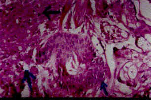
On investigation, hemoglobin was 10 g/dl, total leukocyte count 152,000/cumm, platelet count was 93,000/cumm, normoblast were 20/1000 WBCs and blast cells were 60% of mye-loid series. Bone marrow aspirate also showed marked preponderance of promyelocytes. Fine needle aspiration of nodules showed blast cells. Skin biopsy showed dermal infiltration by monomorphic cells with a thin rim of eosino-philic cytoplasm and large open vesicular nuclei giving an impression of leukemic deposit (Fig. 2). The patient was treated with allopurinol for 14 days and trans retinoic acid for thirty days along with hydroxyurea. There was partial response but within 7 days of completion of therapy, nodules reappeared all over the body. Congenital leukemia is an extremely rare disease and only 25% develop cutaneous deposits. Myeloblastic congenital leukemia is 7 times more common than lymphoid variety but reverse is the usual pattern in childhood. Congenital leukemia has been associated with Down syndrome, Turner syndrome, Klienfelter syndrome but none of these conditions was clinically present in this case. M.K. Dasgupta, Correspondence to:
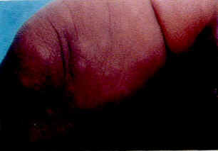
Fig. 1. Photograph showing typical nodular lesions over thigh.
Fig. 2. Biopsy from nodules showing deposit of leukemic cells beside one hair follicle. |
![]()