|
|
|
Indian Pediatr 2020;57:
467-468 |
 |
Serial Computed
Tomography Findings in a Child with Coronavirus Disease
(COVID-19) Pneumonia
|
|
Guiqing He1, Wenjie Sun2,
Jing Wu3 and Jing Cai 4*
1Department of Infectious Diseases and Infectious
Diseases Laboratory, and 4Department of Comprehensive
Medicine, Wenzhou Sixth People’s Hospital, Wenzhou Central
Hospital Medical Group, Wenzhou, China; 2The Second
Affiliated Hospital of Fujian Traditional Chinese
Medical University, Fuzhou, China; and 3Department of
Infectious Diseases,
Huashan Hospital, Fudan University,
Shanghai, China.
Email:
[email protected]
Published online: April 09, 2020;
PII: S097475591600158 |
Novel coronavirus disease (COVID-19) is a highly infectious
disease with its outbreak in China in late 2019 [1]. The novel
coronavirus is reportedly affecting more adults than children
[2,3]. Here, we provide computed tomography (CT) findings in a
typical pediatric case with confirmed COVID-19 infection.
An 11-year-old boy, the close contact of confirmed COVID-19
infected father, presented to hospital with high fever for 10
days. He was confirmed COVID-19 infection by throat swab
specimen test using Realtime RT-polymerase chain reaction
(RT-PCR) method.
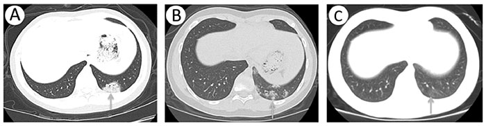 |
| Fig. 1 Chest
computed tomography (CT) scans in an 11-year-old boy
with coronavirus disease-19. a) Chest CT performed on
the day of admission shows patchy ground-glass opacities
in left lower lobe with air bronchogram; b)Follow-up CT
obtained on day 7 shows scattered ground-glass opacities
in left lower lobe which were partly resolved;
c)Follow-up CT obtained on day 14 shows slight sporadic
ground-glass opacities in left lower lobe which have
significantly resolved. |
His symptoms relieved somewhat after interferon a-2b
combined with aerosol therapy in a local hospital. On admission,
arterial blood gas analysis showed a low PaO2of 69.6 mmHg. Chest
CT was performed, which showed patchy ground-glass opacities in
left lower lobe with air bronchogram (Fig.1a). He was diagnosed
as COVID-19 pneumonia. During hospitalization, the child
received recombinant human interferon alpha-2b (rhIFNa2b)
twice-a-day through nebulization combined with Complementary and
alternative medicines. Supportive care including nasal cannula
(maximum oxygen requirement 2L/min) was administered. CT done
one week later (day 7) showed scattered ground-glass opacities
in left lower lobe (Fig. 1b). After two weeks of therapy, only
slight sporadic ground-glass opacities in left lower lobe were
found in repeat chest CT (Fig. 1c). Realtime RT-PCR on two
throat swab specimens was negative for the COVID-19 at 14 weeks,
48 hour apart. The boy made a complete recovery.
This
communication underscores the course of CT findings in COVID-19
pneumonia in a child without any co-morbidity, who improved
after treatment.
Authors’ contributions: GH,JC:
conceptualized the study, collected data, WS: conceptualized the
study, drafted the initial manuscript, and reviewed and JW:
carried out the analyses; all authors reviewed and revised the
manuscript, and approved the final manuscript as submitted.
Competing interests: none stated. Funding: Wenzhou Municipal
Science and Technology Bureau (ZY202004).
REFERENCES
1. Zhu N, Zhang D, Wang W, Li X, Yang
B, Song J, et al. A novel coronavirus from patients with
pneumonia in China, 2019. N Engl J Med. 2020;382:727-33.
2. Cai J, Xu J, Lin D, Yang Z, Xu L, Qu Z, et al. A case series
of children with 2019 novel coronavirus infection: Clinical and
epidemiological features. Clin Infect Dis. 2020 Feb 28 [Online
ahead of print]. Available
from:https://academic.oup.com/cid/advance-article/doi/10.1093/cid/ciaa198/5766430.
Accessed April 2, 2020.
3. Guan WJ, Ni ZY, Hu Y, Liang
WH, Ou CQ, He JX, et al. Clinical characteristics of coronavirus
disease 2019 in China. N Engl J Med. 2020 Feb 28. Available
from: https://www.nejm.org/doi/full/10.1056/NEJMoa2002032.
Accessed April 2, 2020.
|
|
|
 |
|
