|
|
|
Indian Pediatr 2019;56: 433 |
 |
Tinea Faciei Incognito
|
|
Aounallah Amina and Mokni Sana
From the Department of
Dermatology, Farhat Hached Hospital, Tunisia.
Email:
amina_aounallah@yahoo.fr
|
|
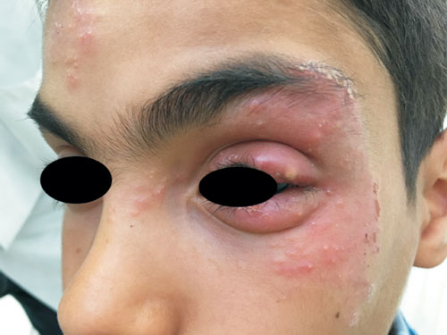
An 11-year-old boy without any medical history was seen by a general
practitioner for inflammatory lesions of the left eyelid, and treated as
eczema with topical corticosteroids (betamethasone) for last one month.
Dermatological examination revealed a squamous and erythematous lesions
strewn with papules and pustules, localized to the left upper and lower
eyelids, with an extension to the periorbital region and forehead (Fig.
1). Mycological examination revealed hyphae, with Microsporum
canis species identified on culture on Sabouraud’s medium. We
diagnosed Tinea faciei incognito was due to Microsporum canis,
and triggered by the local application of corticosteroids. The organism
was most likely transmitted to the patient from his cat. The patient was
advised twice daily applications of ketoconazole cream for 3 weeks, with
which the lesion resolved completely, and mycological examination was
negative.
 |
|
Fig. 1 Pustular erythematous lesion
of the upper and lower left eyelids in Tinea facia incognito.
|
Tinea faciei incognito is often difficult to diagnose
because of the modification of the usual aspect of the dermatophytosis
by the topical corticosteroids. The differential diagnoses for lesion at
this site include contact dermatitis (very itchy, and its elementary
lesion is a vesicle) and blepharitis due to Demodex mites (rosacea-like
dermatitis, folliculitis, and blepharitis). The presence of satellite
pustules and microscopic examination facilitates the diagnosis of Tinea
faciei.
|
|
|
 |
|
