|
|
|
Indian Pediatr 2018;55: 427-428 |
 |
Neonatal Mucormycosis with Gastrointestinal
and Cutaneous involvement
|
|
R Usha Devi 1,
Anitha Balachandran1,
CN Kamalarathnam1
and S Pappathi2
From Departments of 1Neonatology and
2Pathology, Institute of Child Health and Hospital for Children,
Egmore, Chennai, India.
Correspondence to: Dr M Anitha, Assistant Professor,
Department of Neonatology, Institute of Child Health and Hospital for
Children, Egmore, Chennai 600 008, India.
Email: drmanithadm@gmail.com
Received: June 23, 2017;
Initial review: October 09, 2017;
Accepted: January 22, 2018.
|
Background: Mucormycosis of the gastrointestinal tract is a rare
fungal infection of neonates. Case characteristics: 48-hours-old
term neonate presented with intestinal obstruction and perforation. No
significant risk factors were present. Histopathological examination of
the resected gangrenous bowel revealed mucormycosis. Cutaneous
involvement due to systemic spread led to dermal necrosis in toes.
Outcome: Though cutaneous lesions responded promptly to antifungal
therapy, gastrointestinal manifestations required multiple
antifungal therapy for prolonged period apart from surgical debridement.
Message: Precise histopathological diagnosis and early
appropriate therapy can prevent dismal outcomes in neonatal mucormycosis.
Keywords: Gangrene, Intestinal perforation, Necrotizing
enterocolitis, Neonate.
|
|
N
eonatal mucormycosis is a rare, fatal and
opportunistic fungal infection [1,2]. Gastrointestinal mucormycosis is
associated with high mortality, and most cases of cutaneous involvement
need surgical debridement in addition to systemic antifungal drugs [3].
We report a case of neonatal mucormycosis with gastrointestinal and
cutaneous involvement concurrently.
Case Report
A male neonate weighing 2.7 kg delivered at 39 weeks
of gestation after normal transition had passed meconium and was on
direct breast feeds. After 48 hours, he developed signs of intestinal
obstruction and perforation. During laparotomy, two stricturous segments
were noticed in the terminal ileum with friable gangrenous bowel about
20 cm from the ileocecal junction. After excision of stricturous
segments, double barrel ileostomy was done. Thrombocytopenia and
abnormal coagulogram persisted in the neonate even after receiving broad
spectrum antibiotics, vitamin K and fresh frozen plasma transfusions.
Subsequently, cutaneous necrosis was noticed in the great, second and
third toes (Fig. 1). Doppler study revealed normal blood
flow pattern in the involved limb. Biopsy report of the excised bowel
revealed necrotizing enterocolitis with presence of aseptate right
angled fungal hyphae suggestive of mucormycosis (Web
Fig. 1). Urine analysis, renal imaging, eye
examination and echocardiography did not reveal any evidence of fungal
infection. The neonate was treated with Amphotericin B after the biopsy
report. His cutaneous necrosis resolved completely after one week of
Amphotericin B therapy. Ileostomy closure and re-anastamosis was done
during the fourth week. Postoperatively after 72 hours, the infant
deteriorated due to anastamotic leak and the entire small bowel had
formed a cocoon (Web Fig.
1). The abscess was drained and colonic reanastamosis was
attempted during the relaparotomy. Infant continued to have low grade
fever and weight loss despite total parenteral nutrition followed by
enteral feeds. Considering that the gastrointestinal lesion was not
responding to Amphotericin B, Caspofungin was started. Subsequently the
infant became afebrile, started gaining weight and the wound healed
without complications. The infant was discharged on direct breast feeds
after six weeks. During follow-up, the infant was thriving well and
work-up for immunodeficiency states was negative.
 |
|
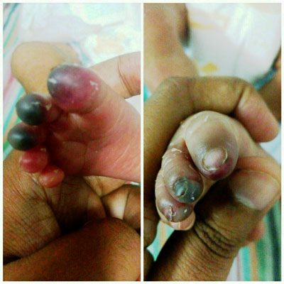
Fig. 1 Cutaneous necrosis of
toes noticed on day 2 of admission (a), and its resolution by 1
week of starting amphotericin B (b).
|
Discussion
Neonatal mucormycosis is rare, fulminant and often
fatal [3]. Among neonates, gastrointestinal tract seems to be
predominantly involved accounting for more than half (54%) of all the
published cases of neonatal mucormycosis [4]. Colon is the most commonly
affected organ among neonates. In our infant, terminal ileum was
involved. The cutaneous lesions in mucormycosis may be caused by an
infection at the primary site or secondary to dissemination from another
site [3]. Dissemination from the gastrointestinal tract could have
resulted in cutaneous mycosis in our neonate.
Due to their angioinvasive nature, these organisms
penetrate through the endothdial cells and in that process can lead to
rapid infarction of tissues. In almost all reported cases of neonates
with gastrointestinal mucormycosis, the organism was identified by
histopathological examination. Isolation of the organism by culture was
possible in only less than half of reported [5]. Mucormycosis is
considered as a variant of necrotizing enterocolitis with similar
clinical presentation. However, neutropenia, absence of pneumatosis
intestinalis and poor response to broad-spectrum antibiotics point
towards mucormycosis [6]. The cutaneous lesion in our case responded
well and healed after one week of systemic antifungal therapy unlike
other cases reported in literature. Though cutaneous necrosis resolved
with short course of Amphotericin B (1 week), gastrointestinal
involvement in our case required prolonged therapy (5 weeks) for
complete recovery. Ischemic necrosis of infected tissues prevents
delivery of leukocytes and antifungal agents to the foci of infection
making the infection extremely difficult to treat with medical therapy
alone [7]. Thus gastrointestinal mucormycosis with severe necrosis of
bowel is associated with high mortality in neonates. Prompt surgical
intervention and institution of appropriate antifungal led to remarkable
improvement and survival in our child preventing the dismal
complications. The initial resection of necrotic bowel probably reduced
the fungal load and improved the response to antifungal therapy in our
case. Delayed anastamosis under the cover of appropriate antifungal
therapy probably helped in better healing. Primary anastamosis is
undesirable as it may lead to extensive gangrene of abdominal wall
following closure.
The median (IQR) age for onset of neonatal
mucormycosis is 12 (8, 18) days [8]. Dhingra, et al. [5] reported
a very early presentation at 24 hours in a neonate with no risk factors,
similar to our case . The risk factors for gastrointestinal mucormycosis
are prematurity, low birth weight, poor nutritional status, diarrhea,
acidosis, hyperglycemia, corticosteroid use, antibiotic adminis-tration,
major surgery, oral or nasogastric tube placement, endotracheal
intubation, indomethacin therapy, asphyxia and contaminated dietary
supplements [9]. Placement of nasogastric tube to the check patency of
esophagus in the delivery room was the only identified risk factor in
our infant. This insertion could have caused local tissue damage and
permitted subsequent early mycotic invasion. Improvement in clinical
status after therapy and normal blood counts did not favour an
immunodeficiency state during the hospital stay. The work-up for immuno-deficiency
done subsequently on follow-up was also normal.
We conclude that the neonates presenting with
clinical features of necrotizing enterocolitis but without pneumatosis
intestinalis, neutropenia and unresponsive to conventional treatment
should arouse suspicion of mucormycosis. Early surgical intervention and
appropriate adequate and prolonged coverage with antifungal therapy can
prevent the dismal outcomes of this treatable condition.
Contributors: RUS,MA: managed the patient;
RUS: reviewed the literature and drafted the initial version of the
manuscript; MA,CNK,SP: contributed to literature review and critically
revised the manuscript. All the authors contributed to drafting of the
manuscript and approved the final version of the manuscript.
Funding: None; Competing interests:
None stated.
References
1. Reimund E, Ramos A. Disseminated neonatal
gastrointestinal mucormycosis: a case report and review of the
literature. Pediatr Pathol. 1994;14:385-9.
2. Sarin YK. Intestinal mucormycosis in a neonate: A
case report and review. J Indian Assoc Pediatr Surg. 2010;15: 98-100.
3. Oh D, Notrica D. Primary cutaneous mucormycosis in
infants and neonates: case report and review of the literature. J
Pediatr Surg. 2002;37:1607-11.
4. Roilides E, Zaoutis TE, Walsh TJ. Invasive
zygomycosis in neonates and children. Clin Microbiol Infect. 2009;15:
50-4.
5. Dhingra KK, Mandal S, Khurana N. Unsuspected
intestinal mucormycosis in a neonate presenting as necrotizing
enterocolitis (NEC). Eur J Pediatr Surg. 2008;18:119-20.
6. Patra S, Chirla D, Kumar N, Vij M, Samal S.
Unsuspected invasive neonatal gastrointestinal mucormycosis: A
clinicopathological study of six cases from a tertiary care hospital. J
Indian Assoc Pediatr Surg. 2012;17:153.
7. Ibrahim AS, Spellberg B, Walsh TJ, Kontoyiannis
DP. Pathogenesis of mucormycosis. Clin Infect Dis. 2012;54:S16-22.
8. Roilides E, Zaoutis TE, Katragkou A, Benjamin DK,
Walsh TJ. Zygomycosis in neonates: an uncommon but life-threatening
infection. Am J Perinatol. 2009; 26:565-73.
9. Vallabhaneni S, Mody RK. Gastrointestinal
mucormycosis in neonates: A review. Curr Fungal Infect Rep. 2015;9:
269-74.
|
|
|
 |
|
