|
|
|
Indian Pediatr 2016;53:
425-426 |
 |
Infantile Bullous Pemphigoid Following
Vaccination
|
|
Kavita Bisherwal, Deepika Pandhi, Archana Singal and
*Sonal Sharma
From Department of Dermatology & STD and *Pathology, University
College of Medical Sciences and Guru Teg Bahadur Hospital, University of
Delhi, Dilshad Garden, Delhi, India.
Correspondence to: Dr Deepika Pandhi, Professor, Department of
Dermatology and STD, University College of Medical Sciences and
associated GTB Hospital, Delhi 110 095, India.
Email:
[email protected]
Received: July 25, 2015;
Initial review: November 09, 2015;
Accepted: February 02, 2016.
|
|
Background: Post-vaccination
infantile bullous pemphigod is a rare presentation. Case
characteristics: A 2-month-old girl presented with widespread bullae,
erosions, necrotic and targetoid lesions over body and mucosae after
vaccination. Histology and direct immunofluorescence (DIF) were
consistent with bullous pemphigoid. Intervention: Clinical
remission with oral steroids and no recurrence with subsequent
vaccination. Message: Continuation of vaccination may not be
contraindicated in infants where bullous pemphigoid onset occurs after
vaccination.
Keywords: Adverse events following
immunization, Infant, Vaccine.
|
|
Bullous pemphigoid is an acquired autoimmune
subepidermal blistering disorder, commonly affecting the elderly
population [1]. Pediatric autoimmune blistering disorders including
bullous pemphigoid are uncommon and have varied manifestations, thereby
leading to difficulty in clinical diagnoses [1]. Pediatric bullous
pemphigoid differs from adult variety by more frequent involvement of
mucous membranes, face, palms, soles, no association with any neoplasm
and rapid resolution with steroids [2].
We report a 2-month-old female infant who presented with
bullous pemphigoid two days after receiving DPT and hepatitis B
vaccination.
Case Report
A 2-month-old girl presented to the dermatology
outpatient department with complaint of fluid filled lesions over limbs,
face, trunk, and groin area of 10 days duration. The lesions initially
appeared over the chest region and extended to involve limbs, palms,
soles, face, abdomen, oral and genital mucosae, and scalp over the next
two days. There was no history of accompanying fever, diarrhea or prior
drug intake, and the child was taking feeds normally. She received first
dose of DPT and hepatitis B vaccines two days prior to onset of lesions.
She was a product of full term normal vaginal delivery born of a non
consanguineous marriage, and there was no history of similar complaints
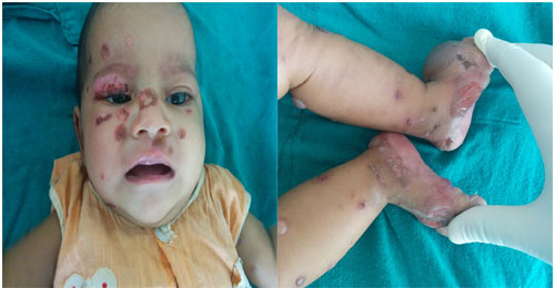
in the family members. Examination revealed multiple tense vesicles and
bullae predominantly over extremities, palms, soles and face. There were
multiple targetoid lesions over trunk and multiple erosions all over
body including scalp, oral and genital mucosae (Fig. 1).
Tzanck smear showed few eosinophils without acantholytic cells and no
organisms were seen on Grams staining. She had been started on
intravenous vancomycin with a provisional diagnosis of bullous impetigo,
with no response. All hematological investigations were unremarkable and
blood culture was negative. A skin biopsy and direct immunofluorescence
(DIF) was performed with differential diagnoses of linear IgA dermatosis
and infantile bullous pemphigoid. Histological examination revealed
subepidermal bulla with dense eosinophilic infiltrate (Web
Fig. 1a) and DIF showed linear deposits of IgG and
C3 and absence of IgA and IgM at dermo-epidermal junction (Web
Fig 1b). With a final diagnosis of infantile
bullous pemphigoid, oral prednisolone therapy was initiated at a dose of
1 mg/kg per day. Rapid improvement with resolution of bullae was
observed after 2 weeks, and the steroids were slowly tapered. The
child’s vaccination was continued as per schedule and no recurrence was
observed with second dose of DPT and hepatitis B vaccine. The child is
in remission at 6 months follow-up.
 |
|
(a)
(b)
Fig. 1 (a) Bullae and crusted erosions over face; and (b)
multiple bullae over lower limbs.
|
Discussion
In pediatric bullous pemphigoid, large tense bullae
are seen predominantly over inner thighs, groin, abdomen, forearms,
axillae, palms and soles and mucous membranes [1]. Infantile bullous
pemphigoid has more widespread clinical lesions with less mucosal
involvement and predominant acral involvement [3,4]; whereas, in
childhood bullous pemphigoid, involvement is severe, less uniform, and
may be localized to genital area [5]. Palmoplantar lesions are
considered as a diagnostic clue of infantile bullous pemphigoid [5].
Schwieger-Briel, et al.
[5] proposed the following minimal diagnostic criteria for infantile
bullous pemphigoid: typical clinical picture (plaques and blisters,
acral distribution) and linear IgG and/or C3 deposition at the basement
membrane in DIF.
The cause of pediatric bullous pemphigoid is unknown
and the possible triggering factors reported include non-specific
maternal antibodies and foreign antigen (infectious agents, drugs,
vaccines) [1,6]. Vaccinations implicated include diphtheria, tetanus,
pertussis, poliomyelitis, influenza, hepatitis B, meningococcal C,
pneumococcus, BCG and rota virus with a latent period of 1 day to 4
weeks between vaccination and onset of disease [1,5,6]. Most cases of
infantile bullous pemphigoid have been described after first dose of
vaccination [5,7]. Vaccination may unmask subclinical bullous pemphigoid
by enhancing an autoimmune response in immunologically predisposed
individuals [4]. It is also
postulated that the inflammation induced in the skin by vaccination –
rather than the vaccines themselves – might generate anti basement
membrane antibodies consequent to disruption of the basement membrane as
no structural similarities have been seen between the implicated
vaccines and basement membrane proteins [8]. This might explain rarity
amongst children despite frequency of vaccinations. Moreover, no
recurrence has been seen with the reintroduction of the vaccination
schedule and continuation of vaccination as per schedule, is recommended
[4].
The clinical and histological differential diagnoses
of bullous pemphigoid include chronic bullous disease of childhood,
dermatitis herpetiformis, bullous lupus erythematosus, epidermolysis
bullosa acquisita (EBA), porphyria and bullous impetigo [5]. EBA is
rarely seen in children, and the histological differentiation is by
presence of neutrophilic infiltrate and IgG deposits only on the dermal
side (base of the blister) in EBA. The prognosis of pediatric bullous
pemphgoid is good with a possibility of spontaneous remission and
infrequent relapses triggered by infections or tapering of
corticosteroids [5]. Steroids (1 to 2 mg/kg) are the first-line
treatment; tapered slowly to prevent rebound [1,5]. Steroid sparing
agents include dapsone, IVIGs, mycophenolate mofetil, erythromycin,
methotrexate-, cyclophosphamide, azathioprine, rituximab or omali-zumab
but their use warrants further investigation [5].
Contributors: All authors contributed to the
acquisition, analysis or interpretation of clinical and histological
(SS) data; and; contributed to the drafting the work or revising it
critically for important intellectual content. All authors approved the
final version.
Funding: None;
Competing interests: None
stated.
References
1. Wu KG, Chou CS, Hsu CL, Lee ML, Chen CJ, Lee DD.
Childhood bullous pemphigoid: A case report and literature review. J
Clin Exp Dermatol Res. 2013;S6:010.
2. Barreau M, Stefan A, Brouard J, Leconte C, Morice
C, Comoz F, et al. Infantile bullous pemphigoid. Ann Dermatol
Venereol. 2012;139:555-8.
3. Waisbourd-Zinman O, Ben-Amitai D, Cohen AD,
Feinmesser M, Mimouni D, Adir- Shani A, et al. Bullous pemphigoid in
infancy: Clinical and epidemiologic characteristics. J Am Acad Dermatol.
2008;58:41-8.
4. Reis-Filho EG, Silva Tde A, Aguirre LH, Reis CM.
Bullous pemphigoid in a 3-month-old infant: case report and literature
review of this dermatosis in childhood. An Bras Dermatol. 2013;88:961-5.
5. Schwieger-Briel A, Moellmann C, Mattulat B, Schauer
F, Kiritsi D, Schmidt E, et al. Bullous pemphigoid in infants:
characteristics, diagnosis and treatment. Orphanet J Rare
Dis. 2014;9:185.
6. Baykal C, Okan G, Sarica R. Childhood bullous
pemphigoid developed after the first vaccination. J Am Acad Dermatol.
2001;44:348-50.
7. Erbagci Z. Childhood Bullous Pemphigoid Following
Hepatitis B Immunization. J Dermatol. 2002;29:781-5.
8. Cohen AD, Shoenfeld Y. Vaccine-induced autoimmunity. J Autoimmun. 1996;9:699-703.
|
|
|
 |
|

