|
|
|
Indian Pediatr 2012;49: 414-416
|
 |
Acute Myeloid Leukemia Presenting as
Obstructive Jaundice
|
|
Binitha Rajeswari, Anu Ninan, Sindhu Nair Prasannakumari*and
Kusumakumary Parukuttyamma
From the Division of Pediatric Oncology and * Division
of Pathology, Regional Cancer Centre, Trivandrum, India.
Correspondence to: Binitha Rajeswari,
Lecturer, Division of Pediatric Oncology, Regional Cancer Centre,
Trivandrum, India.
Email: [email protected]
Received: January 29, 2011;
Initial review: February 24, 2011;
Accepted: August 30, 2011.
|
Jaundice as a presenting feature of pediatric acute myeloid leukemia is
rare. We report two cases of AML who presented with obstructive
jaundice, one with a malignant stricture at the common bile duct and
other with a granulocytic sarcoma obstructing the bile duct. The
prognosis is poor in these patients.
Key words: Acute myeloid leukemia, Granulocytic sarcoma,
obstructive jaundice.
|
|
Obstructive jaundice as the presenting feature
of acute myeloid leukemia (AML) is rare in children. It may be due
to a stricture of the biliary tree or a granulocytic sarcoma
compressing the biliary tree. We report two such cases.
Case Report
Case 1: A one year old female child presented
to us with pancytopenia (hemoglobin 4.5g/dL, WBC count 2100/mm 3,
platelet count 13,000/mm3).
A thorough evaluation including bone marrow study did not reveal any
definite evidence of malignancy. The blood counts normalized in a
month. She presented two months later, with fever, followed by
increasing jaundice, pale stools and abdominal distension. She was
sick, with severe pallor, jaundice, generalized edema and massive
ascites. Hepatosplenomegaly could not be assessed due to the massive
ascites. Laboratory investigations revealed hemoglobin 4.6 g/dL,
platelet count 33000/mm3,
total count 5100/mm3,
serum bilirubin 4.8 mg/dL (conjugated bilirubin 3.6 mg/dL), SGOT 76
IU/L, SGPT 30 IU/L, ALP 263 IU/L and prothrombin time 13 seconds.
Serologies for HIV and HBsAg were negative. Anti HCV titers were not
done. CT scan and ultrasound scan of the abdomen showed a soft
tissue lesion 6×4cm wedged between the pancreas and liver. There was
moderate bilobar intrahepatic biliary radicle dilatation and common
bile duct dilatation upto pancreatic segments. There was bulky
celiac axis, mesenteric and retroperitoneal lymphadenopathy with
moderate ascites and bilateral minimal pleural effusion. Ascitic
fluid cytology showed atypical cells suggestive of leukemia/lymphoma
infiltration. Flow cytometry analysis done on the ascitic fluid
revealed positivity for CD13, CD33, CD117 and CD7 markers,
diagnostic of AML, possibly M5. Bone marrow study was deferred due
to her poor general condition. The patient was started on
subcutaneous cytosine arabinoside. In the following two weeks she
improved with clearing of jaundice, reduction of abdominal
distension and improvement of blood counts. A bone marrow study done
showed 3% blasts with normal hemopoietic elements. Ultrasound scan
of the abdomen showed disappearance of the mass and return of the
biliary channels to normal size. Despite starting chemotherapy with
intravenous cytosine arabinoside and daunorubicin, she developed
sepsis and died.
Case 2: A previously normal 10 month old
female child, presented with history of progressively increasing
jaundice, clay colored stools and high colored urine of 2 months
duration, followed by swellings over both parotid regions and
multiple ecchymotic patches one month later. The child was sick and
malnourished with deep jaundice, pallor, ecchymoses on the face and
generalized lymphadenopathy. There was massive hepatospleno-megaly
with liver palpable 10 cm below the right costal margin, reaching up
to right iliac fossa and spleen palpable 8 cm below the left costal
margin, crossing the midline beyond the umbilicus. Laboratory
investigations showed hemoglobin 11.7 g/dL, platelet count 23000/mm 3,
WBC count 62000/mm3 (Neutrophils
24%, lymphocytes 42%, myelocytes 10%, abnormal cells 24%), serum
bilirubin 38mg/dL (conjugated bilirubin 30.6 mg/dL), SGOT 108 IU/L,
SGPT 50 IU/L, gamma glutamyl transferase 499 U/L and LDH 1132 U.
Serology for HIV and HBsAg were negative. Anti HCV titers were not
done. Peripheral blood smear examination showed 33% peroxidase
positive myeloid blast cells and a diagnosis of AML was made. The
review of the parotid gland biopsy slides also showed infiltration
by peroxidase positive myeloid blasts. Bone marrow studies could not
be done due to the poor general condition of the patient.
Ultrasonography and CT scan of the abdomen showed intrahepatic
biliary radicle dilatation with distension of the gall bladder and
dilatation of the proximal common bile duct. No mass was visualized.
Magnetic resonance cholangiopancreatogram confirmed the above
findings and revealed an obstruction at the level of proximal common
bile duct possibly due to a malignant stricture. There was also
distension of the gall bladder with dilatation of the right and left
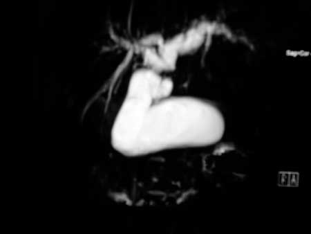
hepatic ducts and cystic duct. (Fig. 1) Chemotherapy
could not be instituted because of the poor general condition of the
patient and she succumbed to her illness.
 |
|
Fig. 1 MRCP showing obstruction at
common bile duct with distension of gall bladder and
dilatation of hepatic ducts.
|
Discussion
Obstructive jaundice as a presenting feature of
pediatric malignancy is rare. Lymphoma and neuroblastoma may present
with biliary obstruction. Rhabdomyosarcoma of the biliary tract may
also occur. Jaundice as a presenting symptom in AML is rare. It can
occur due to drug induced hepatocellular damage, post transfusion
viral hepatitis, infiltration of the liver by the leukemic process
or obstruction of the biliary tract. Obstruction may be due to
granulocytic sarcomas compressing the biliary tree or due to
stricture of the biliary tree. There are very few case reports of
AML presenting as obstructive jaundice, especially in children.
Jaing, et al. [3] have reported a 4 year old boy with
extrahepatic obstruction of the biliary tract in AML. In their
patient, on CT scan of the abdomen, there was a mass lesion at the
pancreatic head associated with biliary dilatation. This patient
responded well to chemotherapy, followed by bone marrow
transplantation and was disease free 15 months after diagnosis.
The granulocytic sarcoma of biliary tree may be
detected radiologically as a stricture or thickening of the biliary
tree[1,2,4,7,8] or as a mass causing extrinsic obstruction of the
biliary tree [3,5,6]. The mass obstructing the biliary tree in AML
is usually a granulocytic sarcoma. This may occur concurrently with
leukemia or may precede the occurrence of leukemia by weeks to
months [5-8]. In our first patient, imaging studies showed a mass
lesion wedged between the pancreas and liver, producing compression
of the biliary channels and so we considered the jaundice to be
obstructive even though the alkaline phosphatase levels were normal.
The mass was a granulocytic sarcoma causing extrinsic compression of
the biliary tree. In our second patient, AML presented as a
stricture of the biliary tree producing obstructive jaundice. In
this scenario, the major differential diagnosis to be considered is
a secondary sclerosing cholangitis which in children could be due to
langerhans cell histiocytosis, immuno-deficiency, sickle cell anemia
or autoimmune diseases. In our patient, since the peripheral smear
was diagnostic of AML, the obstruction was probably due to a
malignant stricture.
Contributors: BR, AN and KP were involved in
patient care. BR and AN collected data and drafted the paper. SNP
was involved in the pathological diagnosis. KP critically analysed
the manuscript. The final manuscript was approved by all authors.
Funding: None; Competing interests:
None stated.
References
1. Mano Y, Yokoyama K, Chen CK, Tsukada Y, Ikeda
Y, Okamoto S. Acute myeloid leukemia presenting with obstructive
jaundice and granulocytic sarcoma of the common bile duct. Rinsho
Ketsueki. 2004;45:1039-43.
2. Rajesh G, Sadasivan S, Hiran KR, Nandakumar R,
Balakrishnan V. Acute myeloid leukemia presenting as obstructive
jaundice. Indian J Gastroenterol. 2006;25: 93-4.
3. Jaing TH, Yang CP, Chang KW, Wang CJ, Chiu CH,
Luo CC. Extrahepatic obstruction of the biliary tract as the
presenting feature of acute myeloid leukemia. J Pediatr
Gastroenterol Nutr. 2001;33:620-2.
4. Lee JY, Lee WS, Jung MK, Jeon SW, Cho CM, Tak
WY, et al. Acute myeloid leukemia presenting as obstructive
jaundice caused by granulocytic sarcoma. Gut Liver. 2007;1:182-5.
5. Matsueda K, Yamamoto H, Doi I. An autopsy case
of granulocytic sarcoma of the porta hepatis causing obstructive
jaundice. J Gastroenterol. 1998; 33:428-33.
6. King DJ, Ewen SWB, Sewell HF, Dawson AA.
Obstructive jaundice-An unusual presentation of granulocytic
sarcoma. Cancer. 2006; 60:114-7.
7. Gonzales-vela MC, Val-bernal JF, Mayorga M,
Cagigal ML, Fernandez F, Mazzora F. Myeloid sarcoma of the
extrahepatic bile ducts presenting as obstructive jaundice. APMIS.
2006; 114:666-8.
8. Sung CO, Ko YH, Park CK, Jang KT, Heo JC.
Isolated biliary granulocytic sarcoma followed by acute myelogeneous
leukemia with multilineage dysplasia: A case report and literature
review. J Korean Med Sci. 2006;21:550-4.
|
|
|
 |
|
