Hypomelanosis of Ito appears to be the third most common neurocutaneous
disease after neurofibro-matosis and tuberous sclerosis. The bizarre,
patterned, hypopigmented macules usually appear during the first year of
life or at birth in sharply demarcated whorls, patches or streaks that
follow the lines of Blaschko. The lesions appear as a negative image of
incontinentia pigmenti. Multiple extracutaneous abnormalities involving
the nervous system, eyes, musculoskeletal, hair, head, face, dental,
cardiovascular, genito-urinary system and other organ anomalies can occur
in most of the patients. We report an interesting case of Hypomelanosis of
Ito with tessellated fundus and polymicrogyria.
 |
|
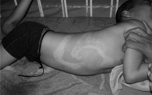
Fig.1 A child with Hypomelanosis of Ito
showing multiple whorled like hypopigmented lesions present over
abdomen, chest and back. |
A 4 years old male child was brought with recurrent
episodes of generalized tonic clonic seizures since 8 months of age. There
was global developmental delay, hypotonia and mental retardation. On
examination, multiple whorled like hypopigmented macules were present over
abdomen, chest, back and both lower limbs arranged along the lines of
Blaschko since birth (Fig. 1). The palms, soles and mucous
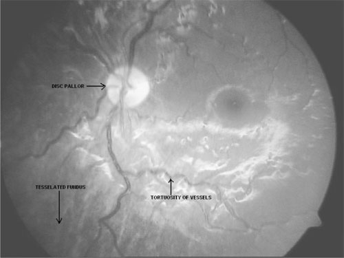
membrane were spared. The fundus showed pale disc, tortous vessels around
the disc, radial patchy streaks of hypopigmentation and tessellated
background (Fig. 2). EEG showed generalized epileptiform
discharges. CT and MRI scan brain showed hemispheric asymmetry with
atrophy of right cerebral hemisphere and ballooning of right lateral
ventricle. MRI brain demonstrated polymicrogyria at right perisylvian and
parietal region, thinning of corpus callosum and periventricular
hyperintense signal.
 |
|
Fig.2 Fundus showing pale disc, tortous
vessels around the disc, radial patchy streaks of hypopig-mentation
and tessellated background. |
Classical tessellated fundus in Hypomelanosis of Ito
has been reported only in two children in literature(1). Also, there are
very few reports about polymicrogyria in Hypomelanosis of Ito(2,3).
Acknowledgment
Prof ML Vasanthakumari, Dr K Mathiarasan, Dr M
Balasubramaniam, Dr S Nataraja Rathinam and Dr V Suba, for their valuable
suggestions and kind cooperation to evaluate this case.
References
1. Hamada T, Saito T, Sugai T, Morita Y. Incontinentia
pigmenti achromians (Ito). Arch Dermatol 1967; 96: 673-676.
2. Steiner J, Adamsbaum C, Desguerres I, Lalande G,
Raynaud F, Ponsot G, et al. Hypomelanosis of Ito and brain
abnormalities: MRI findings and literature review. Pediatr Radiol 1996;
26: 763-768.
3. Liptai Z, Kalmanchey R, Marschalko M, Barsi P. Ito
hypomelanosis (incontinentia pigmenti achro-mians). Orv Hetil 1998; 139:
2587-2591.


