Pre-pubertal gynecomastia is a rare condition in adolescent boys, and may
be idiopathic or may result from excessive estrogen production by adrenal
or testicular tumors, in association with congenital adrenal hyperplasia
or to over expression of aromatase(1). We report a patient presenting with
prepubertal gynecomastia, which on histopatho-logical examination was
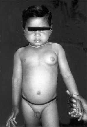
diagnosed as a hamartoma. A 3½ year old male child was brought with the
sole complaint of left sided breast enlargement since 3 years of age. On
examination he had unilateral left sided breast enlargement 5 cm × 5 cm,
non-tender, soft, without any palpable nodule or discharge (Fig.1).
Anthropometric examination was normal for age.
 |
|
Fig. 1 Child with unilateral left sided
gynecomastia. |
Investigations showed serum thyroid stimulating hormone
of 2.9 mU/L, free thyroxine 100nmol/L, leutinising hormone 0.7IU/L,
follicle stimulating hormone 0.8 IU/L, prolactin 2.6 µg/L, oestradiol 20
pmol/L, 17-hydroxy-progesterone 4 nmol/L, dehydro epiandrosterone
sulfate 0.1µmol/L, andro-stenedione 0.2 nmol/L and testosterone 0.2nmol/L
which were all within the normal reference range for his age. Liver and
renal function tests, a-feto protein
and g-hCG were also normal.
Karyotyping showed normal 46XY male. CT scan of the abdomen and pelvis was
normal. USG of the breast showed a well-circumscribed, solid, hypoechoic
mass with posterior acoustic shadowing. CT scan of thorax showed a
unilateral breast tumor, which was well demarcated, and not infiltrating
the chest wall.
Over the next 6 months, his breast swelling increased
to 7 cm × 5 cm and hence a subcutaneous mastectomy was performed.
Histopathological examination showed well capsulated fleshy mass measuring
6×5×5 cm, which was soft in consistency, with yellow islands of fat
tissue. Microscopy showed breast tissue with many cystically dilated ducts
with irregular lumina, lobular cells forming acini and occasional foamy
and myoepithelial cells. Islands of adipose tissue and dense fibrous
tissue were seen. Histopathology thus confirmed hamartomatous fibrocystic
breast mass(2).
In summary, we describe a rare case of a three-year-old
child presenting as pre-pubertal gyne-comastia diagnosed as hamartoma on
excision biopsy. As the underlying pathophysiology of pre pubertal
gynecomastia has not yet been determined, detailed evaluation of such
patients is necessary before surgical intervention is undertaken.
References
1. Braunstein GD. Aromatase and gynecomastia. Endocr
Relat Cancer 1999; 6: 315-324.
2. Deshpande A, Munshi M. Mammary hamartoma: report of two cases
including one in a male breast, and review of the literature. Indian J
Pathol Microbiol 2004; 47: 511-515.


