|
|
|
Indian Pediatr 2009;46: 43 4 |
 |
Subluxation of Lens in Marfan Syndrome |
|
Sumana Datta (Kanjilal) and *Himadri Datta
Department of Pediatrics, Calcutta National Medical
College and Hospital and *Regional Institute of Ophthalmology,
Calcutta, India. E-mail:
[email protected] |
|
A 13-year old boy presented with loss of
vision in both the eyes. On examination the boy had long thin limbs, arm
span (175 cm) exceeded his height (165 cm), ratio being 1.06:1. The lower
segment of his body (87.5 cm) exceeded the upper segment (75 cm), US: LS
was 0.83. He had arachnodactyly of hands and the fingers were
hyperextensible. Steinberg sign was positive i.e., the thumb could be
adducted across the narrow palm. The wrist sign was also positive. The
thoracic cage revealed pectus excavatum and scoliosis was present. Ocular
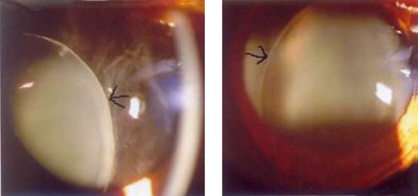
manifestations included myopia and iridodonesis. Slit lamp examination
revealed subluxation of lens in both eyes (Fig.1) Fundus
examination revealed bilateral retinal detachment . Systemic examination
revealed no other abnormalities. Echocardiography revealed mitral
regurgitation. The elder brother has almost similar abnormalities
indicating the hereditary origin.
 |
|
Fig. 1 Subluxation of lens in both eyes. |
Marfan syndrome, caused by mutations in the fibrillin
gene on chromosome 15, presents with abnormalities of the cardiovascular,
musculoske-letal, and ocular systems. Nearsightedness and astigmatism are
common, but farsightedness can also result. Subluxation of lens (ectopia
lentis) in one or both eyes (in 80% of patients) also occurs. In Marfan
syndrome the dislocation is usually super-otemporal (in 75% of cases).
Typically, the zonules that are visible are intact and unbroken, in
contrast to the broken zonules seen in homocystinuria. Some-times eye
problems appear only after weakening of connective tissue has caused
detachment of retina in the second and third decade of life. Early onset
glaucoma can be another complication. Other systemic disorders associated
with ectopia lentis include homocystinuria, where the lens is displaced
inferonasally, Weill-Marchesani syndrome, where the lens is displaced
downwards and forwards and the lens tends to be small and round. In
ectopia lentis et pupillae, an autosomal recessive disorder, both the lens
are displaced in opposite directions. Other causes of ectopia lentis are-
Ehler-Danlos, Sturge-Weber, Crouzon and Klippel-Feil syndromes, oxycephaly
and mandibulo-facial dysostosis.
|
|
|
 |
|
