|
|
|
Indian Pediatr 2009;46: 428-429 |
 |
Congenital Lymphangioma Circumscriptum of the
Vulva |
|
Kamal Aggarwal, Sanjeev Gupta
†, Vijay Kumar Jain and Nisha Marwah*
From the Department of Dermatology, Venereology &
Leprology and Department of Pathology*, Post Graduate Institute of Medical
Sciences and Research, Rohtak, India; and Department of Dermatology,
Venereology & Leprology†, MM Institute of Medical Sciences and Research,
Mullana, Ambala, India.
Correspondence to: Dr Sanjeev Gupta, H. No B-2, Near Shiv
Mandir, MM Medical College Residential Campus, Mullana, District Ambala,
Haryana 133 203. India. E-mail:
[email protected]
Manuscript received: August 29, 2007;
Initial review completed: September 21, 2007;
Revision accepted: May 19, 2008.
|
|
Abstract
Lymphangioma circumscriptum of the vulva is a
disorder of lymphatic channels involving deep dermal tissues. Most of
these cases are confused with genital warts leading to improper
diagnosis and treatment. We present a three years young female child who
had multiple skin colored papular lesions over the genitals. Skin biopsy
revealed features of lymphangioma circumscriptum.
Key words : Child, Lymphangioma, Vulva.
|
|
L
ymphangioma
circumscriptum is a rare benign disorder of no specific etiology involving
the lymphatic channels in the deep dermal and subcutaneous layers(1). It
can occur as either a congenital abnormality or as acquired damage to
previously normal lymphatic channels(2). Axilla, adjacent chest-wall, oral
cavity and tongue are common sites. The vulval presen-tation is
uncommon(1-3). Lymphangioma circumscriptum is characterized by clustered
or diffuse thin walled, translucent vesicles 1 to 5mm in diameter and
filled with clear lymphatic fluid. The vesicles may develop on normal skin
or on top of preexisting papules; hyperkeratosis may sometimes give them a
verrucous appearance.
Case Report
A three year old girl was referred for evaluation of
some eruptions of multiple skin colored papular lesions on the genitalia
since birth. There was neither any preceding history of local trauma nor
any drainage of fluid from the site and of sexual assault too. The site of
involvement had progressively become more extensive in the last four
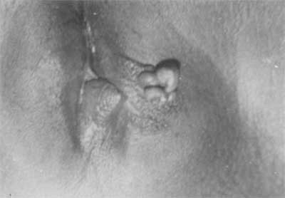
months. On examination, the right labia majora was swollen, hypertrophied
and hyperpigmented. There were multiple verrucous, coalescent papules (Fig.
1). Many of the papules had focal sites of dark red to purple
discoloration (i.e. hemorrhage) within them. No excoriation, crushing or
oozing was observed. The remaining genital and physical examination was
normal. A nontreponemal test for syphilis was non-reactive. The Mantoux
test was negative. Total and differential WBC counts were within normal
limits. Erythrocyte sedimentation rate was 10mm/hr. (Westergren). There
was no evidence of pulmonary tuberculosis in a chest radiograph. In view
of clinical presentation possibility of genital warts, lympha-ngioma
circumscriptum, cystic hygroma was kept. A skin biopsy of a representative
lesion revealed the features of lymphangioma circumscriptum, namely,
dilatation of the lymphatic channels in the papillary dermis with
overlying epidermal acanthosis and hyperkeratosis.
 |
|
Fig.1 Lymphangioma circumscriptum of the
vulva showing verrucous, coalescent papules. |
Discussion
Vulva is an uncommon site for the development of
lymphangiomata. Only 32 cases have been described previously (12
congenital and 20 acquired)(1,3). The youngest age of presentation of
congenital lymphangioma circumscriptum of the vulva, till date, had been
14 years(4). Our case is probably the youngest in terms of age of
presentation of lympha-ngioma of the vulva. Frequent complications of
lymphangioma circumscriptum include swelling of the vulva, pain, recurrent
cellulitis caused by excoriation or spontaneous vesicles, and subsequent
infection of the vulvar area(1). The added compli-cation of psychosexual
dysfunction may lead to cessation of sexual activity. A rare major
compli-cation is lymphangiosarcoma arising at the site of a preexisting
lymphangioma circumscriptum(1).
Treatment modalities include sclerotherapy,
electrocoagulation, liquid nitrogen therapy, carbon dioxide laser therapy,
radiotherapy and surgical excision(2,3,5).
Lymphangioma circumscriptum of the vulva may be
clinically indistinguishable from molluscum contagiosum, genital warts or
tuberculosis verrucosa cutis. Biopsy is essential to confirm the
diagnosis.
References
1. Vlastos AT, Malpica A, Follen M. Lymphangioma
circumscriptum of the vulva: A review of the literature. Obstet Gynaecol
2003; 101: 946-954.
2. Whimster IW. The pathology of lymphangioma
circumscriptum. Br J Dermatol 1976; 94: 473-486.
3. Roy KK, Agarwal R, Agarwal S, Kumar S, Malhotra N,
Gopendru N. Recurrent vulval congenital lymphangioma circumscriptum- a
case report and literature review. Int J Gynae Cancer 2006; 16: 930-934.
4. Bauer BS, Kernahan DA, Hugo NE. Lymph-angioma
circumscriptum - A clinicopathological review. Ann Plast Surg 1981; 7:
318-326.
5. Yildiz F, Atahan IL, Karcaltincaba M, Cengiz M,
Ozyigit G, Aydin A, et al. Radiotherapy in congenital vulvar
Lymphangioma circumscritum. Int J Gynecol Cancer 2008; 18: 556-559.
|
|
|
 |
|
