|
|
Case Reports Indian Pediatrics 2006; 43:437-440 |
||
|
Mucopolysaccharidosis IIIB, Cerebral Vasculopathy and Recurrent Subdural Hematoma |
||
|
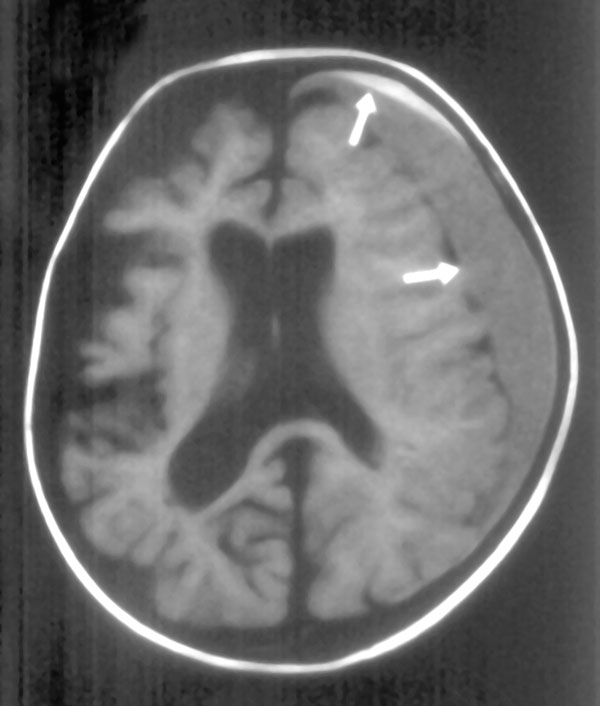
Abstract: Key words: Mucopolysaccharidosis IIIB, Subdural hematoma, vasculopathy. The mucopolysaccharidoses (MPSs) are a family of heritable disorders caused by deficiency of lysosomal enzymes needed to degrade glycosaminoglycans (GAGs). The four-biochemically distinct variants of Sanfilippo disease (MPS IIIA, B, C, and D) are autosomal recessive diseases caused by mutations in one of four genes, which encode enzyme activities required for the lysosomal degradation of heparan sulfate and clinically indistinguishable from each other. The diagnosis is confirmed by analysis in fibroblast for the four enzymes being deficient in different variants of the disease. MPS IIIB (Sanfilippo B syndrome, absence of alpha-N-acetylglucosaminidase: NAGLU) is a rare and progressive disorder characterized by severe central nervous system (CNS) degeneration leading to premature death together with mild somatic features. Mental retardation is severe and progressive. Hepatosplenomegaly, stiff-ness of joints, dwarfism, and skeletal changes are mild or moderate, and corneal clouding is absent(1-4). MPSs are characterized by involvement of multiple organs, including the CNS, liver, spleen, heart, lymph nodes, bone marrow, and blood vessels(5). Though vasculitis is among the causes of recurrent subdural hematoma(5-7), no case of subdural hematoma due to cerebral vasculopathy secondary to MPSs has been reported. To our knowledge, this is the first case of cerebral vasculopathy secondary to MPSs causing recurrent subdural hematoma. Case Report A 3-year-old girl patient clinically a case of MPS was admitted to our clinic with the complaints of vomiting and restlessness. She was the first child of a second-degree consanguineous couple and had problems of delayed speech and walking. Physical examination revealed short stature. Ht 82.5 cm (less than 3rd percentile) and macrocephaly head circumference 54 cm. Her develop-mental mile stones including mental/motor development and linguistic skills were delayed. She had also irritability and walking apraxia. Vital parameters were normal, except bradyarrhythmia (44/min). The examination of other systems was normal. No external injury was detected. Thyroid hormone levels were normal. The enzyme studies revealed a deficiency of NAGLU in plasma. The NAGLU activity was 0.6 µmol/L/hr with a control value of 31 units. These results were obtained from Willink Biochemical Genetics Unit, Regional Screening Unit and Reference Centre for Inherited Metabolic Disease, University of Manchester School of Medicine with number 42452. The marked reduction in this enzyme activity is typical for MPS IIIB(6). Studies for bleeding diathesis, including thrombocyte function tests were normal. Echocardiography and radiographies of skeletal system were normal. Abdominal ultrasonography was relevant with hepatosplenomegaly. In 10 month’s follow-up period, four subdural haematoma attacks have been observed, three of them were left and one of them was right sided (Fig. 1). In magnetic resonance angiography, images of upper parts of circle of Willus and vascular structures in this level were normal. Vascular structures below circle of Willus could not be visualized. Dural biopsy, which was performed while draining the developed subdural haematoma, revealed inflammatory cells with a little number of lymphocytes, polymorphonuclear leukocytes and dominance of eosinophils along the vascular wall. Studies for the other causes of vasculitis e.g., autoimmune diseases, infections were normal. Prednisolone therapy (2 mg/kg/day, in 4 doses) was given for a month. Since then, the patient has been followed for 15 months without any further problems.
Discussion In MPS IIIB neurological problems frequently arise at the end of two years of age. The accumulated mucopolysaccharides are generally found in mononuclear phagocytic cells, endothelial cells, intimal smooth muscle cells, and fibroblasts throughout the body. Accumulation of the substrate in lysosomes lead to degeneration of the CNS with progressive dementia often combined with hyperactivity, sleep disorders, and aggressive behavior. Brain magnetic resonance imaging (MRI) demonstrates white matter abnormalities, cortical atrophy, ventricular enlargement thickening of the diploe, callosal atrophy, basal ganglia involvement, cerebellar changes and dilatation of venous sinuses(6). These findings seem to correspond with the development of pathologic changes in MPS, such as perivascular pits in the white matter observed on slices of the fixed brain(1,5-7). Neurodegeneration is prominent in MPS IIIB(5,7). Vasculitis can be broadly classified into two major categories, namely, primary, referring to those patients with vasculitis isolated primarily to the brain, cord, and their leptomeninges occurring in the absence of recognizable triggers or conditions, and secondary, referring to conditions when co-factors are apparent(8), as being in our patient which secondary to MPS. Besides the other organs, MPSs also cause lesions in vascular structures(5). Subendothelial arterial deposits and allergic vasculitis particularly in the coronary arteries and lesions in the brain may cause ischemia and infarction. It has been known that most of the vasculitis syndromes are related to vascular immunecomplexes. Immune-complexes that form in the presence of excess antigen are soluble. Unless, these complexes, that reached sufficiently high concentrations in the serum are cleaned effectively from the circulation by the mononuclear phagocyte system, they attach vascular wall and start the process of vasculitis with polymorphonuclear, mono-nuclear, and eosinophilic cells(5,9). Similar mechanism is also involved in MPSs. Tissue deposits are accompanied by a mastocytosis. Significant histological features include (a) large quantities of GAGs accompanied by smooth muscle hyperplasia in the intima and media and (b) degenerative changes with mineralization of the internal elastic lamina of arteries and of mucoid material in the media(10). These vessels may be more vulnerable than the vessels of healthy individuals. It seems that the ccrebral vasculopathy in our case is secondary to MPS, and might be reason of recurrent subdural haematoma. No detinitc treatment is available for MPS IIIB patients. Bone marrow transplantation is among the new management approaches in MPS IIIB. The role of a lentiviral vector as the transducing agent of NAGLU cDNA in MPS IIIB fibroblasts has been evaluated(2). Our patient’s 15-month follow-up after steroid therapy had no problems, and this can be an evidence of effectiveness of steroid therapy in vasculopathy that had developed in MPSs patients. Before a conclusive statement about MPS IIIB giving rise to vasculopathy and central hemorrhages, evidence from postmortem brain investigations is needed. Contributors: MA assisted in preparation of the protocol, draft of the manuscript, conceptualized and designed the study, management of patient. SA was involved in supervision and monitoring of laboratory work, collection of the data. NK was responsible for preparation of the protocol, draft of the manuscript, conceptualized and designed the study, management of patient. NA participated in histological evaluation of dural biopsy. Funding: None. Competing interests: None stated. | ||
|
References | ||
|
|
![]()