|
|
Case Reports Indian Pediatrics 2002; 39:495-497 |
||
|
Unilateral Porencephaly |
||
|
S. Bhagyabati Devi
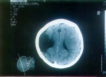
Porencephaly is a pseudocyst secondary to an infarct or other destructive cerebral lesion(1). It is a rare condition probably caused by vascular occlusion resulting from an insult during fetal development or an injury occurring later in life. It is often associated with various ophthalmic and neurologic signs including visual field defects, abnormal pupillary responses, optic nerve hypoplasia, decreased vision, nystagmus, strabismus, hemi-in-attention, seizures and mental deficiencies(2). There are two types of porencephaly: (i) Type I is generally due to an antepartum intraparenchymal hemorrhage; and (ii) Type II lesions are usually due to developmental anomalies. prognosis of each type generally depends on the extent of the lesion(3). We report a case of unilateral porencephaly. Case Report A 15-year-old boy presented with repeated attacks of intractable convulsions since the age of 8 months. Various seizure patterns were noticed in the last 15 years such as drop attacks, complex partial seizures and generalized tonic clonic seizures. The symptoms were worsening gradually since the age of 18 months. Antenatal history was uneventful. Birth history revealed presence of fetal distress with an Apgar score of 3/10 at 1 minute, 7/10 at 20 minutes with birth weight of 2500 g. There was no congenital abnormality detected clinically at birth. Parents noticed less movement of right extremities with inability to stand and walk at the age of 12 months. The patient was examined by a pediatrician and was diagnosed as right sided hemiplegia with hemiatrophy. The fundus examination was shown to be normal. Random blood sugar and X-rays of chest and skull were normal. The child was treated by conventional anticonvulsant drugs till the age of 13 years since the age of 8 months. He also had an intellectual impairment with delayed milestone. Due to lack of proper diagnosis he was put on multiple combinations of anticonvulsant drugs till the age of 13 years. CT scan of brain done at the age of 13 years showed a porencephalic cyst in left cerebral hemisphere (Fig. 1). EEG showed increased theta and delta waves taken over the porencephalic cyst. CT scan of brain taken at the age of 15 years showed the same picture. Discussion Large cystic lesions within the substance of the brain have been found in patients with congenital hemiplegia, mental retardation and other chronic neurological handicaps from the nineteenth century onwards. Heschl who had earlier proposed the term "Porencephaly" in 1868 for such lesions, suggested that they might result from reduction of the blood supply to a localized area of the brain. Their location and the frequent angiographic finding of occluded anterior or middle cerebral arteries strongly support an ischemic aetiology. In many cases the cysts develop prenatally. Their frequent association with polymicrogyria suggests that in these cases both originate before the sixth month of gestation. In other cases, origin at the time of delivery seems likely from circumstantial evidence and rarely, the history suggests a postnatal onset when the sudden development of an acute hemiplegia in a previously normal child is followed later by the demonstration of a porencephalic cyst in the appropriate situation(3). Polyporencephaly is another rare condi-tion, in which both hemispheres are replaced by multiple cystic spaces. The affected infants are usually abnormal at birth with seizures, hypertonia, irritability and an abnormal cry. Sometimes the fetal movements have been reduced and there is often a history of neonatal asphyxia, which may be the result rather than the cause of the cerebral abnormality. The prognosis is very poor, most children dying within a few weeks or months(3).
Fig. 1. CT scan at the age of 13 years showing the porencephalic cyst in left cerebral hemisphere. Antenatal diagnosis of porencephaly by Ultrasonography is possible from the third trimester in case of congenital origin(4). USG, CT scan and MRI can detect the cyst which were previously missed on an air encephalo-graphy since porencephalic cyst did not communicate with the ventricular system. EEG taken over porencephalic cysts are characterized by an increased theta and delta bands in the areas surrounding the lesion sites identified by CT(5). Contributors: All the authors were responsible for managing the patient, reviewing the literature and drafting the manuscript. SBD will act as the guarantor for the paper Funding: None. Competing interests: None stated.
| ||
| References | ||
|
![]()